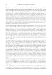
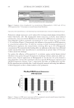
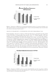
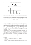
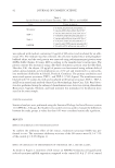
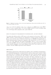
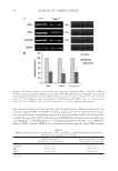
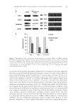
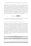
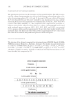
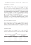
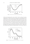
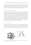
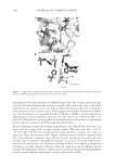
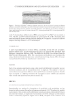
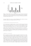
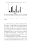
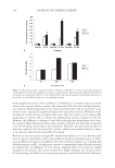
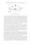
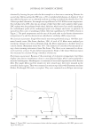
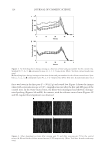
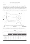
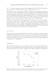
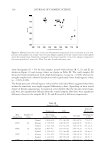
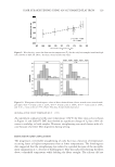
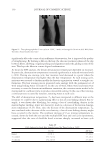
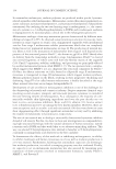
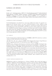
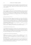
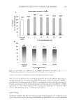
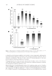
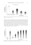
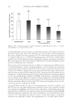
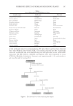
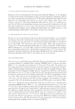
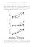
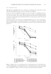
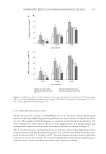
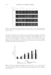
JOURNAL OF COSMETIC SCIENCE 84 INFLUENCE OF HEXAPEPTIDE-11 ON STRESSED-INDUCED PREMATURELY SENESCENT FIBROBLAST CELLS Premature cellular senescence can be induced by treating normal human dermal fi bro- blasts with H2O2 (26). The hexapeptide delays senescence in H2O2 stress-induced prematurely senescent dermal fi broblasts as measured by ATM protein expression in a dose-dependent fashion at peptide concentrations of 1.0% or more, but not at levels 0.1% and below (Figure 3). Interestingly, the infl uence of the peptide on ATM expression at concentrations greater than 1% does not continue to show increasing suppression of ATM protein expression, but instead shows a leveling effect up to 2% of hexapeptide concentration. The reasons for this leveling effect are unknown at this time, but additional studies reported here were typically run at concentrations no greater than 1.0% because of these initial dose fi ndings. To examine the infl uence of Hexapeptide-11 on intrinsic aging, normal human dermal fi broblasts were grown through 24 population doublings without additional added oxi- dative stress. Topical application of Hexapeptide-11 for the last 18 cycles showed a dose-dependent, statistically signifi cant reduction of both ATM protein expression and SA-β-Gal expression, Figures 4 and 5, respectively. The effect on ATM protein and SA- β-Gal expression was reversible after 1 week of peptide removal indicating that infl uence of the hexapeptide on these cellular markers is not permanent. Figure 3. Changes in ATM protein expression in H2O2 prematurely stressed normal human dermal fi bro- blasts treated with various concentrations of Hexapeptide-11. Figure 2. Summary of ratio of medians for two concentrations of Hexapeptide-11, 0.001% and 1.0% for ATM expression after 24 h treatment on normal human dermal fi broblasts.

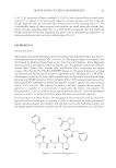
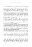
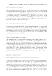
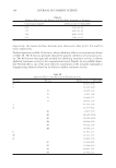
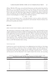
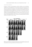
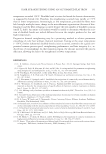
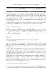
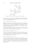
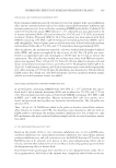
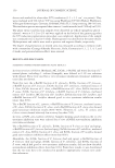
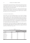
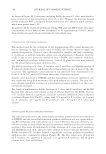
MODULATION OF CELLULAR SENESCENCE 85 INFLUENCE OF HEXAPEPTIDE-11 ON PREMATURELY SENESCENT DERMAL PAPILLAE CELLS Dermal papillae cells grow at the base of the hair shaft and are responsible for the growth of new hair fi bers in anagen phase hair growth cycles (27). Dermal papillae cells from balding and non-balding individuals have been grown ex vivo (5). It has been demon- strated that dermal papillae cells taken from balding areas of the scalp show a higher level of senescence cellular subpopulations compared to dermal papillae from non-balding ar- eas. For the purposes of this study, an in vitro senescence testing model was developed using UV to push dermal papillae cells into premature senescence. Dermal papillae cells were exposed to 20 mJ/cm2 UVB for a period of time previously determined to not be cytotoxic to the cells, but known to elicit expression of measureable quantities of SA- β-Gal. After the irradiation, the cells were incubated for 48 h with various concentrations of the Hexapeptide-11, after which SA-β-Gal activity was determined. Exposure of dermal Figure 4. Expression of ATM protein in normal dermal fi broblasts aged for 22 population doubling cycles and treated for the last 18 cycles with Hexapeptide-11. Data for the same fi broblasts then tested one week after removal of the peptide from the media. Figure 5. Expression of SA-β-Gal protein in normal dermal fi broblasts aged for 22 population doubling cycles and treated for the last 18 cycles with Hexapeptide-11. Data for the same fi broblasts then tested one week after removal of the peptide from the media.
Purchased for the exclusive use of nofirst nolast (unknown)
From: SCC Media Library & Resource Center (library.scconline.org)