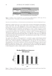
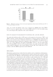
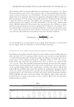
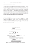
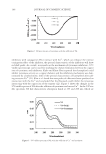
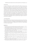
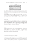
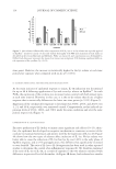
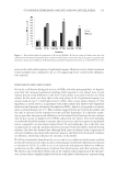
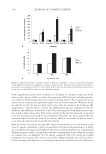
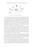
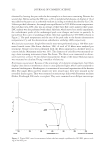
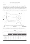
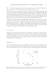
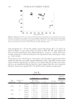
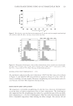
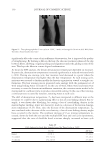
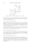
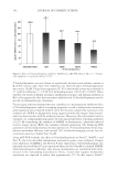
JOURNAL OF COSMETIC SCIENCE 86 papillae cells to UV radiation causes an increase in the expression of SA-β-Gal indicating the cells are expressing biochemical signatures of premature senescence (Figure 6). Treatment with Hexapeptide-11 shows a dose-dependent, statistically signifi cant decline in SA-β-Gal indicating reduced senescence at the 0.5% and 1.0% treatment levels. CONCLUSION Senescence, whether replicative or stress-induced, can be detected in both fi broblasts and dermal papillae cells through analysis of ATM protein expression and SA-β-Gal activity. A hexapeptide originally isolated from S. cerevisiae fermentation lysates and later synthe- sized at high purity when applied to both intrinsically and extrinsically aged fi broblasts in vitro was found to reduce gene expression of ATM and SA-β-Gal proteins in a dose- dependent fashion. Application of Hexapeptide-11 reduced ATM protein expression in fi broblasts that have undergone SIPS using H2O2 and those grown through intrinsic ag- ing cycles. SA-β-Gal activity was reduced in intrinsically aged fi broblasts exposed to Hexapeptide-11 as well. A method of using UV light to induce SIPS in dermal papillae was also developed. It was shown that Hexapeptide-11 also decreased SA-β-Gal activity in UV-irradiated dermal papillae cells. This suggests that the hexapeptide can delay senescence in a second dermal cell line. The infl uence of Hexapeptide-11 on senescence-associated aging appears to be broadly applicable and reversible. To date, this may represent the fi rst known peptide to possess these attributes. The impact of these fi ndings is being further substantiated on ex vivo models. The results of these fi ndings will be reported at a later date. REFERENCES (1) J. A. Knight, The Biochemistry of aging, Adv. Clin. Chem., 35, 1–62 (2000). (2) L. Hayfl ick and P.S. Moorhead, The serial cultivation of human diploid cell strains, Exp. Cell Res., 25, 585–621 (1961). (3) L. Hayfl ick, The limited in vitro lifetime of human diploid cell strains, Exp. Cell Res., 37, 614–636 (1965). Figure 6. Analysis of SA-β-Gal expression in UV-induced prematurely senescent human dermal papillae cells compared against nonirradiated cells.

MODULATION OF CELLULAR SENESCENCE 87 (4) E. Sikora, T. Arendt, M. Bennett, and M. Narita, The impact of cellular senescence signature on ageing research, Ageing Res. Rev., 37, 146–152 (2011). (5) A. W. Bahta, N. Farjo, B. Farjo, and M.P. Philpott, Premature senescence of balding dermal papilla cells in vitro is associated with p16(INK4a) expression, J. Invest. Dermatol., 128, 1088–1094 (2008). (6) F. Debacq-Chainiaux, C. Borlon, T. Pascal, V. Royer, F. Eliaers, N. Ninane, G. Carrard, B. Friguet, F. de Longueville, S. Boffe, J. Remacle, and O. Toussaint, Repeated exposure of human skin fi broblasts to UVB at subcytotoxic level triggers premature senescence through the TGF-β1 signaling pathway, J. Cell Sci., 118, 743–758 (2004). (7) J. Chen, S. Patschan, and M.S. Goligorsky, Stressed-induced premature senescence of endothelial cells, J. Nephrol., 21, 337–344 (2008). (8) D.J Baker, T. Wijshake, T. Tchkonia, N.K. LeBrasseur, B.G. Childs, B. van de Sluis, J.L. Kirkland, and J.M. van Deursen, Clearance of p16Ink4a-positive senescent cells delays ageing-associated disorders, Nature, 479, 232–236 (2011). (9) U. Herbig, W. A. Jobling, B. P. Chen, D. J. Chen, and J. M. Sedivy, Telomere shortening triggers senescence of human cells through a pathway involving ATM, p53, and p21(CIP1), but not p16(INK4a), Mol. Cell, 14, 501–513 (2004). (10) Y. Shiloh and M. B. Kastan, ATM: Genome stability, neuronal development, and cancer cross paths, Adv. Cancer Res., 83, 209–254 (2001). (11) H. Zhan, T. Suzuki, K. Aizawa, K. Miyagawa, and R. Nagai, Ataxia telangiectasia mutated (ATM)- mediated DNA damage response in oxidative stress-induced vascular endothelial cell senescence, J. Biol. Chem., 285, 29662–29670 (2010). (12) M.B. Kastan and D. S. Lim, The many substrates and functions of ATM, Nature Rev. Mol. Cell Biol., 1, 179–186 (2000). (13) G. P. Dimri, X. Lee, G. Basile, M. Acosta, G. Scott, C. Roskelley, E. E. Medrano, M. Linskens, I. Rubeli, O. Pereira-Smith, M. Peacocke, and J. Campisi, A biomarker that identifi es senescent human cells in culture and in aging skin in vivo, Proc. Natl. Acad. Sci., 92, 9363–9367 (1995). (14) D. J. Schlemm, M. J. Crowe, R. B. McNeill, A. E. Stanley, and S. J. Keller, Medicinal yeast extracts, Cell Stress Chaperones, 4, 171–176 (1999). (15) M. J Crowe, R. B. McNeill, D. J. Schlemm, D. G. Greenhalgh, and S. J. Keller, Topical application of yeast extract accelerates the wound healing of diabetic mice, J. Burn Care Rehabil., 20, 155–162(1999). (16) J. M. Liptak, An overview of the topical management of wounds, Aust. Vet., 75, 408–413 (1997). (17) S. J. Keller, R. H. Levin, and J. Fang, Isolation and characterization of a tissue respiratory factor from baker’s yeast, J. Cell Biol., 119, 1005–1008 (1991). (18) J. P. Bentley, T. K. Hunt, J. B. Weiss, C. M. Taylor, A. N. Hanson, G. H. Davies, and B. J. Halliday, Peptides from live yeast cell derivatives stimulate wound healing, Arch. Surg., 125, 641–646 (1990). (19) W. Goodson, D. Hohn, T. K. Hunt, and D. Y. K. Leung, Augmentation of some aspects of wound heal- ing by a skin respiratory factor, J. Surg. Res., 21, 125–129 (1976). (20) O. E. Sorensen, J. B. Cowland, K. Theilgaard-Monch, L. Liu, T. Ganz, and N. Borregaard, Wound heal- ing and expression of antimicrobial peptides/polypeptides in human keratinocytes, a consequence of common growth factors, J. Immunol., 170, 5583–5589 (2003). (21) S. O. Canapp, J. P. Farese, G. S. Schultz, S. Gowda, A. M. Ishak, S. F. Swaim, J. Vangilder, L. Lee- Ambrose, and F. G. Martin, The effect of topical tripeptide-copper complex on healing of ischemic open wounds, Vet. Surg., 32, 515–523 (2003). (22) V. Frei, E. Perrier, I. Orly, A. Huc, C. Augustin, and O. Damour, Activation of fi broblast metabolism in a dermal and skin equivalent model: A screening test for activity of peptides, Int. J. Cosmet. Sci., 20, 159–173 (1998). (23) K. Katayama, J. Armendariz-Borunda, R. Raghow, A. H. Kang, and J. M. Seyer, A pentapeptide from type I procollagen promotes extracellular matrix production, J. Biol. Chem., 268, 9941–9944 (1993). (24) M. P. Lupo and A. L. Cole, Cosmeceutical peptides, Dermatol. Ther., 20, 343–349 (2007). (25) Gruber JV and Holtz R, Examining the genomic infl uence of skin antioxidants in vitro, Mediators Infl amm., 2010, 1–10 (2010). (26) S. Zdanov, J. Remacle, and O. Toussaint, Establishment of H2O2-induced premature senescence in hu- man fi broblasts concomitant with increased cellular production of H2O2, Ann NY Acad Sci., 1067, 210–216 (2006). (27) D. J. Tobin, Ex vivo organ culture of human hair follicles: A model epithelial-neuroectodermal-mesenchymal interaction system, Methods Mol. Biol., 695, 213–227 (2011).
Purchased for the exclusive use of nofirst nolast (unknown)
From: SCC Media Library & Resource Center (library.scconline.org)