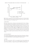
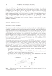
JOURNAL OF COSMETIC SCIENCE 14 cells migrated from the original attachment site. The cells were cultured in DMEM/FBS in air/5% CO2 and temperature of 37oC. Cell treatment. To determine the cytotoxicity of the extract, we transferred the cell suspen- sion (not exceeding eight passages) from a 175-cm2 fl ask into a 96-well plate (1 × 10⁴ cells per well). After 24 h incubation, cells were exposed to 50–200 μg/ml extract for 24 h. The control cells were cultured in DMEM without extract. The viability of cells was determined by 2,3-bis(2-methoxy-4-nitro-5-sulfophenyl)-5-[(phenylamino)carbonyl]- 2H-tetrazolium hydroxide (XTT Boehringer Mannheim, Mannheim, Germany) assay (17,18). Briefl y, following incubation, the old medium was replaced with 200 μl serum- free DMEM and 50 μl XTT-labeling mixture was added. The samples were further incu- bated for 4 h. The intensity of dye was measured with a spectrophotometer at 490 nm. The fraction of viable cells was calculated by subtracting the optical density fraction of treated cells from the untreated cells. These determinations were performed in triplicate. EFFECTS OF THE EXTRACT TO H2O2-DAMAGED FIBROBLASTS A suspension of fi broblasts from the primary cultures (not exceeding eight passages) was transferred from a 175-cm2 fl ask into a 24-well plate (1 × 10⁵ cells per well) and then incubated at 37°C under 5% CO2 for 24 h. After treatment with 200 μg/ml tamarind seed coat extract, cells were treated with various concentrations (100–1000 μM) of H2O2 to induce oxidative stress in fi broblast (19). The control cells were not exposed to H2O2 or the extract. The viability of cells was determined by XTT assay. The cytotoxicity of H2O2 on cells was quantifi ed and expressed as percentage of cell damage relative to 0% damage calculated from the number of control cells. The study was performed in tripli- cate. DETERMINATION OF PREVENTIVE EFFECTS OF THE EXTRACT TO UVA-INDUCED CELL ALTERATIONS UVA irradiation. The UVA source was generated by a Honle F-lamp with a H-1 band- pass fi lter (320–400 nm) and the output intensity measured by a meter with UVA probe (Honle, Gräfelfi ng, Germany). A suspension of fi broblasts from the primary cultures (not exceeding eight passages) was transferred from a 175-cm2 fl ask into 12-well plates (2 × 10⁵ cells per well). There were three treatment groups: (1) no extract, no UVA irradiation (control group) (2) no extract plus UVA irradiation and (3) extract pretreatment plus UVA irradiation group. For group 3 cells, tamarind seed coat extract was added directly to the medium (200 μg/ml) and all the groups were similarly incubated for 24 h (37°C/5% CO2) after which they were 80–90% confl uent. To avoid UV being absorbed or creating adjuncts, all media were replaced by phosphate-buffered saline (PBS without extract). Some of the plates (groups 2 and 3) were UVA irradiated (40 J/cm2) after which the PBS in all the plates was replaced by culture media. After this, the cells of each group were studied as follows: Cell viability: After 24 h of irradiation, the XTT assay was used to assess cell viability. The optical density of the control group was adjusted to 100%.

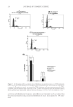
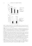
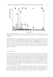
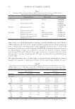
EFFECTS OF TAMARIND SEED COAT EXTRACT ON HUMAN SKIN 15 Cell-cycle analysis: After 72 h of irradiation, the proportions of cells at cell-cycle stages G1, S, and G2 were determined by the DNA stain, propidium iodide (PI Sigma-Aldrich). Cells were detached by trypsinization and washed twice with PBS containing 2 mM EDTA (Sigma-Aldrich). The cells were fi xed overnight with absolute ethanol (−20°C) and then stained with a solution containing 10 μg/ml PI, 10 μg/ml RNAse (GIBCO/ Invitrogen Corporation), and 2 mM EDTA in PBS. After 20 min incubation at room temperature in the dark, fl uorescent cells were sorted in a fl ow cytometry system equipped with a 488-nm argon laser (model FACScalibur, Becton Dickinson, Franklin Lakes, NJ). Cell-cycle data were analyzed using CellQuest Pro software. Total glutathione content: After 6, 24, 48, and 72 h of irradiation, cells were collected and lysed with T-PER lysis buffer (Pierce Biotechnology, Rockford, IL) for 20 min, and the resultant samples were centrifuged (5000g/5 min) to collect the supernatant. Total glu- tathione (GSH/GSSG) used was a commercial kit (Dojindo Molecular Technology, Ku- mamoto, Japan). To each well of 96-well plates, 20 μl enzyme solution, 140 μl coenzyme solution, and 20 μl of either GSH standard solution or the test supernatant were incu- bated at 37oC for 10 min. 5-5’-Dithio-bis(2-nitrobenzoic acid) solution (20 μl) was then added, and the samples were further incubated at 37oC for 20–40 min. Absorbance was measure at 415 nm on a microplate reader (Spectra Count PerkinElmer, Waltham, MA), and GSH content read off a calibration curve. MMP-1 and type I procollagen content: An enzyme-linked immunosorbent assay was used to quantitate human MMP-1, pro- and active forms, and newly synthesized type I procol- lagen in fi broblast cell culture supernatants. After 24–72 h of irradiation, cell-free super- natants were collected and stored at −80°C until used. The amount of MMP-1 and type I procollagen was measure by using a commercial human MMP-1 EIA kit (RayBiotech, Norcross, Germany) and a commercial human procollagen type I C-peptide EIA kit (Ta- kara Bio, Shiga, Japan), respectively. The levels of MMP-1 and type I procollagen were normalized against a standard dose–response curve based on the absorption at 450 nm using a microplate reader. The experiments were performed in triplicate. STATISTICAL ANALYSIS All quantitative data were expressed as means of samples for each treatment. Student’s unpaired t-test was used for comparison between two groups. p 0.05 was considered signifi cant. RESULTS AND DISCUSSION CHARACTERISTICS OF THE TAMARIND SEED COAT EXTRACT AND TOTAL PHENOLIC CONTENT The crude extract from tamarind seed coat was light-brown powder. The percent yield of the extract obtained was 22.0 ± 0.8% w/w. As determined by Folin–Ciocalteu assay, the amount of the phenolic compounds was 58.0 ± 0.3 g gallic acid equivalents per 100 g extract.
Purchased for the exclusive use of nofirst nolast (unknown)
From: SCC Media Library & Resource Center (library.scconline.org)