JOURNAL OF COSMETIC SCIENCE 36 (3) K. R. Millington and J. S. Church, The photodegradation of wool keratin II. Proposed mechanisms involving cystine, J Photochem Photobiol B., 39, 204–212 (1997). (4) R. H. Bradley, I. L. Clackson, and D. E. Sykes, UV ozone modifi cation of wool fi bre surfaces, Appl Surf Sci, 72, 143–147 (1993). (5) D. Yilmazer and M. Kanik, Bleaching of wool with sodium borohydride, J Eng Fiber Fabr., 4, 45–50 (2009). (6) J. M. Dyer, J. Plowman, G. Krsinic, S. Deb-Choudhury, H. Koehn, K. Millington, et al., Proteomic evaluation and location of UVB-induced photo-oxidation in wool, J Photochem Photobiol B., 98, 118–127 (2010). (7) J. M. Dyer, S. Bringans, and W. G. Bryson, Characterisation of photo-oxidation products within pho- toyellowed wool proteins: Tryptophan and tyrosine derived chromophores, Photochem Photobiol Sci., 5, 698–706 (2006). (8) B. Mahltig, F. Audenaert, and H. Bottcher, Hydrophobic silica sol coatings on textiles—The infl uence of solvent and sol concentration, J Sol-Gel Sci Technol., 34, 103–109 (2005). (9) Technical release: Luzchem exposure standard: LES-UVA-01 (2004) Available from: http://www. luzchem.com/handbook/LESUVA011.pdf. (10) Technical release: Luzchem exposure standard: LES-UVB-01 (2004) Available from: http://www. luzchem.com/handbook/LESUVB011.pdf. (11) Technical release: Luzchem exposure standard: LES-420-01 (2004) Available from: http://www.luzchem. com/handbook/LES420011.pdf. (12) H. Thomas, A. Conrads, K. H. Phan, M. van de Löcht, and H. Zahn, In vitro reconstitution of wool intermediate fi laments, Int J Biol Macromol., 8, 258–264 (1986). (13) N. R. Parker, J. F. Jamie, M. J. Davies, and R. J. W. Truscott, Protein-bound kynurenine is a photosen- sitizer of oxidative damage, Free Radic Biol Med., 37, 1479–1489 (2004). (14) A. Pirie, Formation of N′-formylkynurenine in proteins from lens and other sources by exposure to sunlight, Biochem J., 125, 203–208 (1971). (15) T. Gensch, J. Hendriks, and K. J. Hellingwerf, Tryptophan fl uorescence monitors structural changes accompanying signalling state formation in the photocycle of photoactive yellow protein, Photochem Photobiol Sci., 3, 531–536 (2004). (16) J. Lee, N. Koo, and D. B. Min, Reactive oxygen species, aging, and antioxidative nutraceuticals, Compr Rev Food Sci Food Saf., 3, 21–33 (2004). (17) C. Wei, B. Song, J. Yuan, Z. Feng, G. Jia, and C. Li, Luminescence and Raman spectroscopic studies on the damage of tryptophan, histidine and carnosine by singlet oxygen, J Photochem Photobiol A Chem., 189, 39–45 (2007). (18) J. A. Maclaren and B. Milligan, “The Structure and Composition of Wool,” In Wool Science—The Chem- ical Reactivity of the Wool Fibre (Science Press, Marrickville, Australia, 1981), pp. 1–18. (19) R. C. Marshal, D. F. G. Orwin, and J. M. Gillespie, Structure and biochemistry of mammalian hard keratin, Electron Microsc Rev., 4, 47–83 (1991).

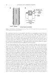
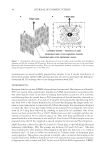
J. Cosmet. Sci., 65, 37–48 (January/February 2014) 37 Preliminary analysis of the distribution of water in human hair by small-angle neutron scattering YASH KAMATH, N. SANJEEVA MURTHY, and RAM RAMAPRASAD, Kamath Consulting Inc., Monmouth Junction, NJ (Y.K.), Center for Biomaterials Rutgers University, Piscataway, NJ (N.S.M), and TRI/Princeton, Princeton, NJ (R.R.). Accepted for publication November 17, 2013. Synopsis Diffusion and distribution of water in hair can reveal the internal structure of hair that determines the pen- etration of various products used to treat hair. The distribution of water into different morphological compo- nents in unmodifi ed hair, cuticle-free hair, and hair saturated with oil at various levels of humidity was examined using small-angle neutron scattering (SANS) by substituting water with deuterium oxide (D2O). Infrared spectroscopy was used to follow hydrogen–deuterium exchange. Water present in hair gives basically two types of responses in SANS: (i) interference patterns, and (ii) central diffuse scattering (CDS) around the beam stop. The amount of water in the matrix between the intermediate fi laments that gives rise to interfer- ence patterns remained essentially constant over the 50–98% humidity range without swelling this region of the fi ber extensively. This observation suggests that a signifi cant fraction of water in the hair, which contrib- utes to the CDS, is likely located in a different morphological region of hair that is more like pores in a fi brous structure, which leads to signifi cant additional swelling of the fi ber. Comparison of the scattering of hair treated with oil shows that soybean oil, which diffuses less into hair, allows more water into hair than coconut oil. These preliminary results illustrate the utility of SANS for evaluating and understanding the diffusion of deuterated liquids into different morphological structures in hair. INTRODUCTION Hair is a part of skin, a protective organ of most mammals. It consists of mainly keratin, a structural protein that is also a major constituent of hoofs, nails, and horns. Although hair has a molecularly complex hierarchical structure (Fig. 1) (1), three distinct parts can be identifi ed in the cross sections of human hair fi bers: the cuticle, the cortex, and the medulla (2). The cuticle sheath, which is the outermost protective layer, consists of sev- eral layers of cuticle cells arranged like shingles on a roof. The cuticle envelopes the cor- tex, which is made of cortical cells that comprise the major part of the α-keratin in hair. Cells in both the cuticle and the cortex are held together by cell membrane complexes. Cells in the cortex occur as an assemblage of macrofi brils with rodlike intermediate fi laments Address all correspondence to Yash Kamath at yashkamath@verizon.net.
Purchased for the exclusive use of nofirst nolast (unknown)
From: SCC Media Library & Resource Center (library.scconline.org)