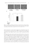
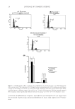
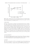
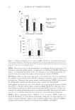
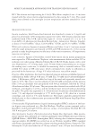
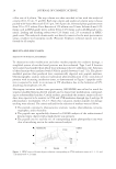
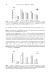
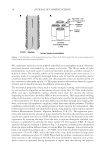
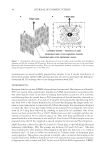
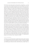
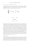
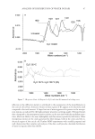
JOURNAL OF COSMETIC SCIENCE 40 measurements on several carefully prepared hair samples to see if similar distribution of water exists in hair. Infrared (IR) spectroscopy data are used to investigate the hydrogen– deuterium (H–D) exchange that occurs during saturation with D2O. EXPERIMENTAL European dark brown hair (EDBH) obtained from International Hair Importers (Glendale, NY) was used in these experiments. Samples for SANS measurements were prepared by fi rst removing the water in the hair by keeping them under vacuum for 24 h, and then equilibrating them for several days in D2O atmosphere of desired humidity obtained us- ing saturated salt solutions. A second set of samples were prepared by equilibrating them fi rst with D2O at the desired humidity for 24 h and then keeping the sample under vac- uum at room temperature to remove free D2O from the sample. For experiments designed to study the effect of oil, hair soaked with oil was prepared by treating the two tresses with oil (0.5 ml/g hair) followed by combing to spread the oil uniformly. One of the tresses was heat-treated for 90 s at 180°C. Both tresses were left overnight and then im- mersed in hexane for 30 s to remove surface oil. They were then equilibrated to the re- quired humidity with solutions of salt and D2O. Cuticle-free (CF) hair samples were also prepared using the CF hair supplied as special samples by International Hair Importers. SANS data were collected on the CG-2 and CG-3 beam lines at the Oak Ridge National Laboratory (Oak Ridge, TN). The q-range was between 0.05 and 0.025 nm−1. The data were normalized to tress thickness. Hair aligned in the form of a thin tress (~2–3 mm in thickness and ~15 mm in width) was mounted on U-frames made of aluminum plate using superglue. These plates were placed in airtight cells to maintain the samples at the required humidity. Different humidity levels were maintained using saturated solutions of appropriate salts in water and D2O. In the case of D2O the RH is expressed as %RHD. Figure 3. (A) Schematic illustration of the distribution of water in a fi ber with crystalline and amorphous domains and (B) the resulting SANS pattern. Water in the interlamellar spaces gives rise to the meridional (fi ber-axis) and off-meridional scattering. Water in the longitudinal channels contributes to the scattering along the equator (perpendicular to the fi ber-axis).

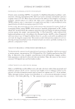
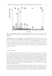
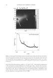
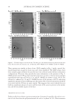
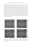
ANALYSIS OF DISTRIBUTION OF WATER IN HAIR 41 Attenuated total refl ectance (ATR) spectra were collected using Nicolet 6700 FTIR (Thermo Electron Corporation, Madison WI). The respective hair sample was placed on the ATR crystal. The spectral region from 900 to 4000 cm−1 was scanned. Spectra were analyzed using OMNIC software (Thermo Electron Corporation). RESULTS AND DISCUSSION INTERNAL STRUCTURE OF HAIR An example of the two-dimensional (2D) SANS pattern is shown in Fig. 4A. There is a key difference between Fig. 4A and Fig. 2 that was used for illustrating SANS. The dominant feature in Fig. 2 is attributed to the water in the interlamellar spaces, and is therefore aligned close to the fi ber-axis (off-meridional). In contrast, the dominant feature in Fig. 4A is attributed to the water in the channels along the fi ber-axis, and therefore occurs along the equator. We attribute the equatorial peak to the water in the spaces be- tween the IFs. Only half of the scattering pattern was obtained so as to record the second maximum. This second peak is signifi cantly weaker than the fi rst. A one-dimensional intensity scan (I vs. q) perpendicular to the fi ber-axes obtained from this image is plotted in Fig. 4B. Here, q is the scattering vector defi ned as q = 4π sinθ/λ (1) where λ is the wavelength of the neutrons (0.478 nm) and 2θ is the scattering angle. The spacing (d) between the scattering entities is calculated by Bragg’s equation d = 2π /q (2) The large scattering intensity, shown in dark (or red) color in Fig. 4A, arises from both the contrast due to D2O and due to the H–D exchange in the amine and amide groups in the hair structure. As shown using the IR data in a later section, the water taken up initially is strongly adsorbed, or even exchanged between the amine and amide functionality of the protein. The changes in the SANS intensity with the D2O concentration in hair can be understood by considering the changes in the contrast in the dry and the D2O-wet hair. Because hydro- gen has negative scattering length, there is a small negative contrast in the dry samples. As shown later in the section on CF hair, this negative contrast gives rise to a SANS interfer- ence peak even in dry samples. With infusion of some D2O, this contrast disappears at 6 %RHD, and the scattered intensity approaches zero. As the hair continues to absorb more D2O, contrast reaches large positive values and the intensity increases considerably. The interference peaks at q = 0.07 Å−1, the dumbbell-like pattern, and a weaker peak at 0.13 Å−1 are evidence of D2O diffusing into the matrix between the IFs, as well as into the IFs. The d-spacing corresponding to the peak at q = 0.07 Å−1 in Fig. 4B is 95 Å. This is consistent with 75 Å size of the IF (Fig. 1B). The second weak peak at q ~0.13 Å−1 (d = 48 Å) has contributions from the internal structure and from the lipids in keratinous tis- sue (12,13). Although it was claimed that the presence of this ring could be used to di- agnose breast cancer (14), this was later refuted (13). Consistent with these latter papers, our study data show that the 0.13 Å−1 peak is a composite of the second order of refl ection from IF assembly and from the lipids (to be published).
Purchased for the exclusive use of nofirst nolast (unknown)
From: SCC Media Library & Resource Center (library.scconline.org)