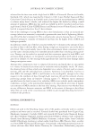
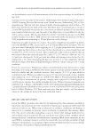
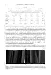
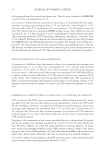
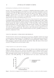
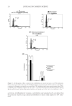
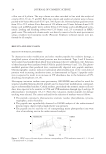
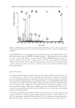
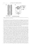
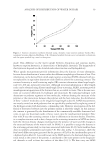
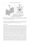
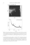
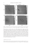
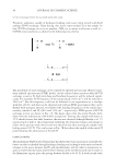
JOURNAL OF COSMETIC SCIENCE 42 Given that both the interference peaks (q ~0.07 and 0.13 Å−1) arise from the organization of the intermediate and protofi laments, we would expect that as the hair takes up mois- ture and swells at the macroscopic level, there would be swelling of the structures at the level of IFs as well. This was indeed observed as the interference peaks (“dumbbells” spots in Fig. 4A) move inwards, corresponding to an increase in the d-spacing from 95 to 101 Å as the humidity is increased from 32 to 92 %RH. Figure 4. (A) A typical 2D small-angle neutron scattering from a hair sample. The dark spot is the shadow of beam stop that blocks the main beam. Side bar shows the intensity scale. The large arrow indicates the direction of alignment of hair fi bers. The two small arrows indicate the two interference peaks. These data were obtained from a hair tress equilibrated at 75 %RHD (B) one-dimensional (1D) intensity scan as a func- tion of scattering vector obtained from the 2D image by summing the intensity along a slice perpendicular the fi ber-axis.

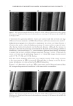
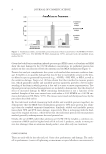
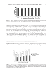
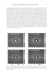
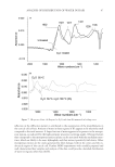
ANALYSIS OF DISTRIBUTION OF WATER IN HAIR 43 One observation in this study is that the scattering intensity corresponding to q = 0.07 and 0.13 Å−1 did not change signifi cantly with the amount of water (D2O) in the sample after the fi rst hydration up to 50 %RH. This suggests that the fraction of water in the vicinity of the IFs in the cortical cells remains relatively constant over the 50–95% humidity range. This was also refl ected in the increase in the d-spacings. The increase in the d-spacings cor- responds to a swelling of ~13%. However, Fig. 3 shows that the amount of water absorbed reaches 12% at 50 %RH, and then accelerates to reach value of ~25% as the humidity is increased to 95%. As amount of absorbed water increases with humidity, it shows that the additional water is present in regions other than the IF assemblies. The amount of water in these regions is expected to be different from that bound to the amide groups in the protein matrix. These assertions are supported by depolarization thermal-current (DTC) study in which ice-like water was reported at 19% water content (15). This water does not have the rotational properties of free water but has the polarization properties of the water molecules in the liquid state. Note that the 19% water content at which this new water peak appears in the DTC study is about the same value at which the amount of water accelerates in sorp- tion isotherm experiments (Fig. 2). The central diffuse scattering (CDS) observed near the beam stop suggests that water could be present in mesoporous (void structures between 100 and 1000 nm in size) regions in hair. Preliminary results in fact show a decrease in this in- tensity in hair exposed to D2O, perhaps because of contrast matching. More than one struc- tural feature contributes to the CDS (16). Important among these are the refraction from the macroscopic surfaces (hair) and microscopic surfaces (internal structure) (10), microvoids within the hair, and fi brils. Because the surface scattering and refraction from surfaces ap- pear in the form of elongated streaks (10), absence of such elongated streaks along the equa- tor allows us to attribute the CDS to internal structures and microvoids within hair. Although water sorption data can be used to determine the pore volume distribution in hair, it will not be accurate, because water swells the hair as it is absorbed, thus continu- ally changing the pore size. Mercury intrusion porosimetry is commonly used for porosity determination. In this method, mercury is forced into the void structure of the fi ber sub- strate under very high pressures (17). This method is generally not used for organic fi bers because high pressures used deform the pore structure in the fi ber. The standard method for determining the pore size distribution in solid substrates such as fi bers is nitrogen adsorption at low temperatures, using Kelvin equation to convert the sorption data into pore volume (18). Gas adsorption measurements indeed show that pores are present in hair, and their distribution changes during various hair treatments (19). The presence of pores is confi rmed by recent detailed electron microscopy studies (20), atomic force mi- croscopy studies (21), and optical measurements (22). Pores in the medullary features form a small fraction of the bulk of hair. Thus, water present in the keratinous structure gives rise to the interference peaks, and water present elsewhere, e.g., longitudinal pores, perhaps in the cortex and the cuticle, contributes to the CDS. SANS OF CUTICLE-FREE HAIR To determine if cuticular sheath is the locus of larger fraction of water in hair, we repeated the scattering experiments with CF hair. Samples of CF hair were prepared in the same way as regular hair as described earlier. Moisture-free samples of CF and regular EDBH were fi rst prepared by keeping them under vacuum in the presence of phosphorous pent- oxide. SANS patterns of the dry (no H2O or D2O) CF-free hair are shown in Fig. 5A.
Purchased for the exclusive use of nofirst nolast (unknown)
From: SCC Media Library & Resource Center (library.scconline.org)