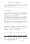
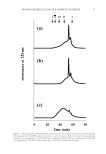
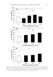
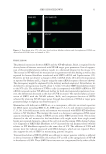
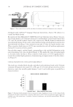
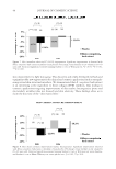
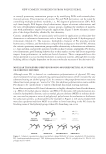
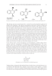
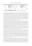
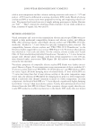
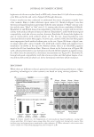
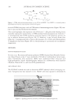
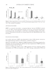
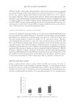
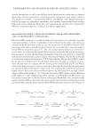
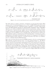
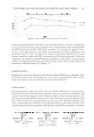
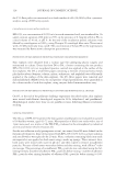
JOURNAL OF COSMETIC SCIENCE 108 Sebum is constituted of oily components such as triglycerides and wax esters. Those are digested by Malassezia strains that converts them into free fatty acids such as oleic acid. This particular metabolite induces scalp fl aking, pruritus, and infl ammation (5,6). How- ever, the relation between free fatty acids and infl ammation remains unexplained. Malassezia fungus naturally belongs to a healthy skin microbiota. Under particular condi- tions, their biology is dysregulated and consequently may contribute to various skin disorders (7,8). For the scalp, specifi c strains of Malassezia have been identifi ed and linked with dandruff and/or SD, namely M. furfur, M. globose, and M. restricta (9,10). They pos- sess a high lipase activity, since their survival is linked to their host’s lipids (10). Oleic acid resulting from Malassezia activity induces a skin infl ammatory response. Toll- like receptor 2 (TLR2) promotes infl ammatory response after binding to the fatty acid, and consequently releasing human beta-defensins (hBD) (11,12). These defensins are upregu- lated in the presence of Malassezia (13). In particular, hBD2 and hBD3 induce adaptive immune responses from keratinocyte which produce pro-infl ammatory cytokines. The resulting situation of those factors induces a disturbance of the scalp stratum corneum (SC) promoting dandruff. The epidermal turnover is dramatically increased which leads to the accumulation of parakeratotic cells with pyknotic nuclei (condensed chromatin) in the upper layers of the epidermis (14). This increased cell proliferation leads to a decreased cell differentiation rate. All together these factors disturb the SC structure. Native from North America, Epilobium angustifolium has been used as a medicinal plant. Folk medicine indicates use of the fi reweed juice to soothe skin irritation and burns. MATERIAL AND METHODS PLANT MATERIAL AND REAGENTS The fi reweed fl ower/leaf/stem aqueous extract (E. angustifolium extract) used in this study is rich in oenothein B (0.12–0.36%), an ellagitannin possessing anti-infl ammatory and antioxidant activity. IN VITRO LIPID SYNTHESIS INHIBITION IN SEBOCYTES Primary human sebocytes were obtained from a 66-year-old female volunteer, presenting a normal body mass index of 23.4. Cells were seeded in 96-black-well/clear-bottom plates, in sebocyte medium. Confl uent cells (fourth passage) were treated with various concentrations of E. angustifolium extract (0.0125%, 0.05%, and 0.1%) for 3 days. The diacylglycerol acyltransferase (DGAT) inhibitor A922500 (2 μM) was used as a control. Intracellular lipid accumulation in cells was evaluated with Nile red staining. Relative fl uorescence intensity was measured at ex/em 540 nm/620 nm (no cutoff) for total lipids and 485 nm/555 nm (515 cutoff) for neutral lipids. EX VIVO MALASSEZIA-INDUCED ALTERATIONS OF SKIN MORPHOLOGY AND IMMUNE MARKERS IN SKIN EXPLANTS Full thickness skin explants were obtained from a 47-year-old Caucasian woman. A 1:1:1 mixture of M. furfur, M. globosa, and M. restricta was prepared, for a total of 12.4 × 10⁶

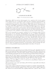
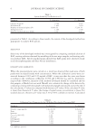
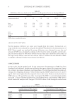
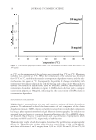
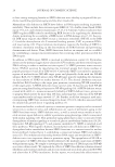
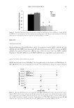
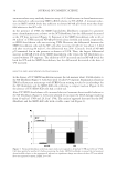
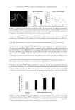
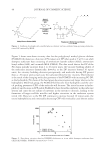
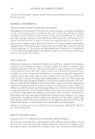
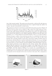
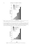
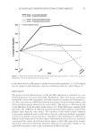
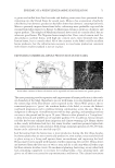
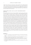
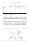
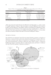
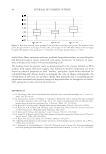
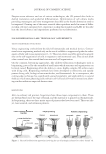
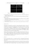
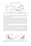
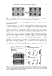
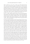
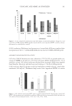
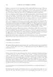
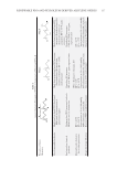
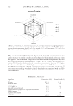
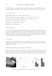
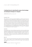
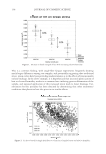
BATTLE AGAINST DANDRUFF 109 CFU/ml. On D0, a fi lter paper disk presoaked in 90 μl of the yeast mixture was applied (or not) for 24 h at the surface of skin explants, for a fi nal concentration of 1.1 × 10⁶ CFU/cm2. On D1, D2, and D4, explants were treated (or not) with a solution of 1.5% E. angustifo- lium extract (2μl/cm2). On D5, explants were collected. Observation of skin general mor- phology by optical microscopy was performed following staining of paraffi nized sections according to Masson’s trichrome Goldner variant. Immunostaining of hBD2 and hBD3 as well as TLR2 was done using antibodies specifi c for each proteins whose presence was subsequently revealed with secondary FITC-conjugate antibodies giving a green fl uores- cence. Cells nuclei were stained with propidium iodide. CLINICAL EVALUATION OF E. ANGUSTIFOLIUM EXTRACT A total of 24 volunteers (men and women, 20–61 years of age) with dandruff and greasy hair participated in the study. Following a 15 days wash-out period using a neutral shampoo, participants were instructed to wash their hair and scalp every 3 days for 30 days, with the same neutral shampoo to which was added 1.5% E. angustifolium extract. The pres- ence of dandruff was estimated on a scale of 0–5 by a trained technician, on D0, D3, D9, D15, and D30 (before shampooing). For this evaluation, the head was divided into four equal parts with a comb. The fi nal grade for dandruff presence on scalp was calculated as the mean of all four head part scores. Adherent and nonadherent dandruff were estimated separately. The presence of sebum on scalp was measured with the Sebumeter® SM 810 (Courage + Khazaka, Cologne, Germany) on D0, D15, and D30. Macrophotographs of scalp were additionally taken in standardized conditions on one preselected zone, at D0, D3, and D30. A self-assessment form was also fi lled out by all subjects on D30 to subjec- tively evaluate the properties of the study product (effi cacy, tolerance, and future use). RESULTS AND DISCUSSION Using a normal human sebocyte culture model and Nile red staining, the effect of E. angustifolium extract on lipid synthesis is presented in Figure 1, as percentages of relative fl uorescence units in comparison with DGAT (taken as 100%), a known inhibitor of lipid synthesis. As can be seen, E. angustifolium extract dose dependently inhibited sebum pro- duction. A substantial reduction by -43% was achieved with a 0.1% concentration of Figure 1. Epilobium angustifolium extract inhibits lipid synthesis in sebocytes.
Purchased for the exclusive use of nofirst nolast (unknown)
From: SCC Media Library & Resource Center (library.scconline.org)