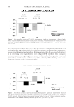
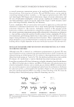
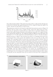
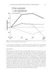
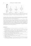
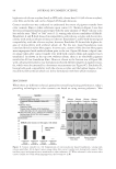
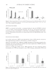
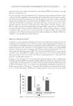
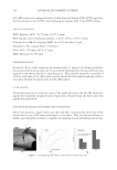
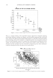
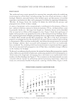
SIRT6 KNOCKDOWN 33 (15) R. D. Ley, Photorepair of pyrimidine dimers in the epidermis of the marsupial Monodelphis domestica, Photochem. Photobiol., 40, 141–143 (1984). (16) J.-P. Etchegaray, L. Zhong, and R. Mostoslavsky, The histone deacetylase SIRT6: At the crossroads between epigenetics, metabolism and disease, Curr. Top. Med. Chem., 13, 2991–3000 (2013). (17) T. L. A. Kawahara, E. Michishita, A. S. Adler, M. Damian, E. Berber, M. Lin, R. A. McCord, K. C. L. Ongaigul, L. D. Boxer, H. Y. Chang, and K. F. Chua, SIRT6 links histone H3 lysine 9 deacetylation to NF-κB-dependent gene expression and organismal life span, Cell, 136, 62–74 (2009). (18) G. Natoli, When sirtuins and NF-κB collide, Cell, 136, 19–21 (2009). (19) Y. Kanfi , S. Naiman, G. Amir, Y. Peshi, G. Zinman, L. Nahum, Z. Bar-Joseph, and H. Y. Cohen, The sirtuin SIRT6 regulates lifespan in male mice, Nature, 483, 218–221 (2012). (20) R. Mostoslavsky, DNA repair, insulin signaling and sirtuins: At the crossroads between cancer and aging, Front. Biosci., 13, 6966–6990 (2008). (21) H. Pan, D. Guan, X. Liu, J. Li, L. Wang, J. Wu, J. Zhou, W. Zhang, R. Ren, W. Zhang, Y. Li, J. Yang, Y. Hao, T. Yuan, G. Yuan, H. Wang, Z. Ju, Z. Mao, J. Li, J. Qu, F. Tang, and G.-H. Liu, SIRT6 safe- guards human mesenchymal stem cells from oxidative stress by coactivating NRF2, Cell Res., 26, 190– 205 (2016). (22) Y. Baohua and L. Li, Effects of SIRT6 silencing on collagen metabolism in human dermal fi broblasts, Cell Biol. Int., 36, 105–108 (2012). (23) K. Wischermann, S. Popp, S. Moshir, K. Scharffetter-Kochanek, M. Wlaschek, F. de Gruijl, W. Hartschuh, R. Greinert, B. Volkmer, A. Faust, A. Rapp, P. Schmezer, and P. Boukamp, UVA radiation causes DNA strand breaks, chromosomal aberrrations and tumorigenic transformation in HaCaT skin keratinocytes, Oncogene, 27, 4269–4280 (2008). (24) L. Zhong, A. D’Urso, D. Toiber, C. Sebastian, R. E. Henry, D. D. Vadysirisack, A. Guimaraes, B. Marinelli, J. D. Wikstrom, T. Nir, C. B. Clish, B. Vaitheesvaran, O. Iliopoulos, I. Kurland, Y. Dor, R. Weissleder, O. S. Shirihai, L. W. Ellisen, J. M. Espinosa, and R. Mostoslavsky, The histone deacetylase Sirt6 regu- lates glucose homeostasis via HIF-1α, Cell, 140, 280–293 (2012).

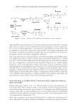
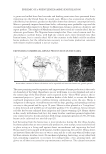
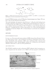
J. Cosmet. Sci., 68, 35–41 ( January/February 2017) 35 Collagen XVIII: A key interfacial component of the skin architecture ISABELLE BONNET, LARA JOBEILI, SÉBATIEN CADAU., NICOLAS BERTHÉLÉMY., AUDREY PIERROT, CARINE TEDESCHI, VINCENT BARDEY, GUILLAUME FARGIER, DELPHINE RIVAL, CHRISTINE JEANMAIRE, CATHERINE BONNAUD-ROSAYE, WENDY CHAN, MANASI CHAVAN, VALÉRIE ANDRÉ-FREI, HASSAN ZAHOUANI, and PATRICIA ROUSSELLE, BASF Beauty Care Solutions France, Lyon, France (B.I., C.S., B.N., P.A., T.C., B.V., F.G., R.D., J.C., B.C., A.V.), BASF Corporation, Tarrytown, NY 10591 (C.W., C.M.), Tribology and System Dynamics Laboratory’s, Ecole Centrale Lyon, Ecully, France (Z.H.), and Laboratoire de Biologie Tissulaire et Ingénierie Thérapeutique, CNRS, Université Lyon 1, Lyon, France (L.J., R.P.). INTRODUCTION Collagen XVIII belongs to the multiplexins, known as extracellular matrix proteins that contain multiple triple-helix domains (collagenous domains) interrupted by noncol- lagenous domains. Besides, as collagen XVIII is a basement membrane (BM) heparan sulfate proteoglycan, it holds the structural properties of both collagen and proteoglycan. This collagen is expressed ubiquitously in various BM structures throughout the body. In skin and compared to other collagen types, collagen XVIII displays the broadest repar- tition as it could be synthetized by keratinocytes, endothelial cells, epithelial cells of the sweat glands and hair follicles stem cells, and adipocytes (1). However, the complete physiological role of collagen XVIII is not fully understood, even if its localization and ultrastructural organization reveal that it is an important component of all BM molecular networks present in skin (2). Understanding its expression modulation with age is of great interest for cosmetic research and could provide new strategies to counteract the loss of tissue structure and cohesion seen during aging of epidermis, dermis, hypodermis, and scalp. Address all correspondence to Valerie Andre-Frei at valerie.andre-frei@basf.com.
Purchased for the exclusive use of nofirst nolast (unknown)
From: SCC Media Library & Resource Center (library.scconline.org)