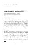
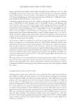
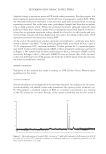
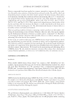
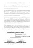
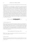
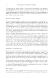
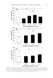
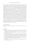
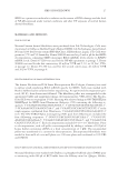
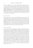
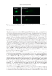
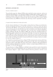
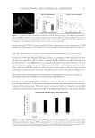
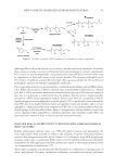
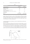
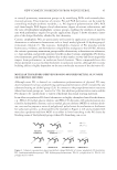
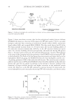
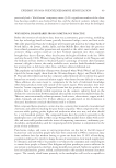
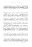
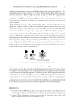
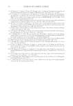
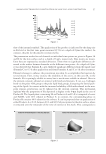
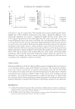
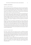
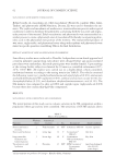
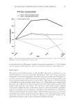
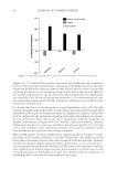
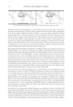
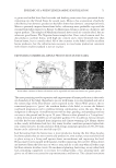
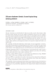
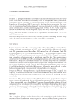
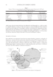
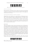
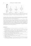
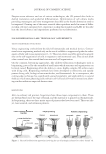
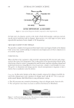
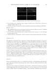
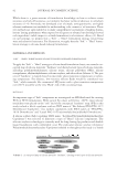
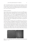
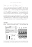
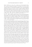
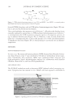
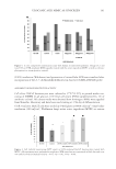
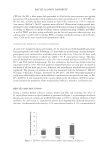
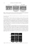
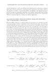
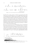
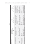
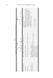
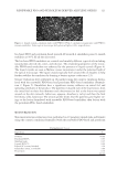
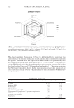
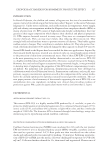
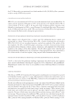
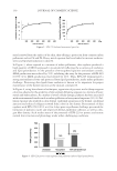
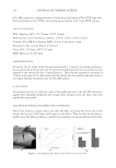
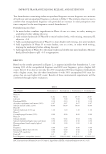
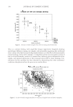
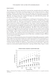
JOURNAL OF COSMETIC SCIENCE 36 METHODS AND RESULTS SPECIFIC ANTIBODY DESIGN To specifi cally target the collagen XVIII portion involved in tissue structure, and not its endostatin domain (Figure 1), we designed an affi nity-purifi ed rabbit antibody against its two collagenic domains: COL1 and COL 5. This antibody was used for collagen XVIII immuno- localization on skin biopsies of different ages and for the design of an enzyme-linked immunosorbent assay (ELISA) on keratinocytes allowing cosmetic ingredient screening. COLLAGEN XVIII EXPRESSION DURING SKIN AGING At fi rst immuno-labelling of 12 skin samples coming from face lifting was performed to localize and quantify this protein in the skin structure (n = 6 for 8–15 years old and for 30–69 years old). Briefl y, 7-μm cryosections were fi xed using cold methanol/acetone during 10 min and unspecifi c sites were blocked using 10% normal goat serum diluted in phosphate- buffered saline (PBS) at room temperature. Then sections were incubated within the primary antibody and after two washes in PBS, the sections were incubated with Cy3- labelled anti-rabbit antibody and counterstained with Dapi. Sections were examined using a Zeiss LSM 700 confocal microscope and the acquisition was realized using the Z-stack mode of the Zen program. Quantifi cation data are presented as mean values and standard error of the mean from at least three measures performed on biological triplicate samples for each skin samples (Figure 2). As compared to young skin specimens, collagen XVIII staining was less intense and homog- enous. In a 60 years old skin sample, heterogeneity of the lamina densa labeling was observed as well as zones where the lamina densa starts to duplicate and detach from the dermal epidermal. Quantifi cation allowed to evidence a strong collagen XVIII decrease as skin ages (-43.5% for older group). The age decrease of collagen XVIII was confi rmed using quantitative reverse transcription polymerase chain reaction (qRT-PCR) study on keratinocytes extracted from 50 abdominal skin biopsies (Figure 2, right), which evidenced that the proteomic decrease occurs Figure 1. Collagen XVIII structure (NC: non collagenic domains, COL: collagenic domains, : antigenic area).

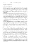
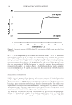
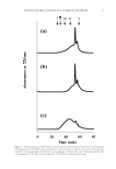
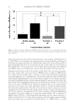
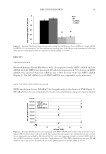
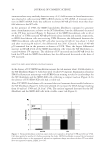
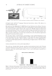
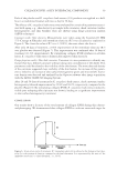
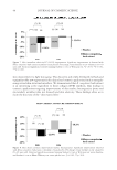
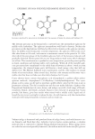
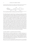
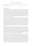
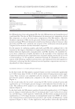
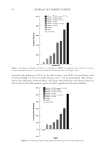
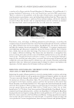
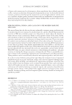
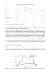
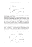
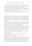
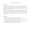
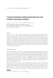
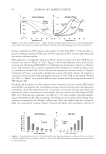
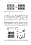
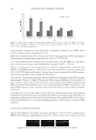
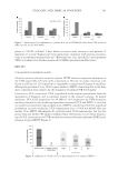
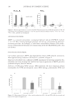
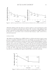
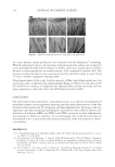
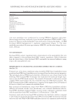
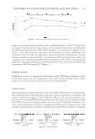
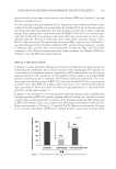
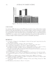
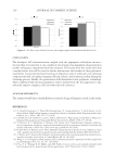
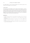
COLLAGEN XVIII: A KEY INTERFACIAL COMPONENT 37 mainly through COL18A1 gene regulation. Gene expression was also reduced after UVB irradiation, highlighting the implication of this target in photoaging (data not shown). NEW ELISA DEVELOPMENT TO SELECT ACTIVE INGREDIENT PROMOTING COL XVIII SYNTHESIS In order to restore the collagen XVIII age decrease, a screening test was developed to iden- tify the best ingredient able to induce collagen XVIII synthesis in adult keratinocytes. For this purpose, a new ELISA test was designed on keratinocytes extracted from a 36 years old skin specimen using the specifi c affi nity-purifi ed rabbit antibody previously described. After culture and ELISA protocol optimizations, several vegetal extracts were evaluated and Khaya senegalensis bark extract was identifi ed as one of the most effi cient (Figure 3). NEW NONCONTACT DEVICE DESIGN FOR MECHANICAL PROPERTIES EVALUATION IN VITRO ON RECONSTRUCTED SKINS TREATED BY K. SENEGALENSIS BARK EXTRACT In order to evaluate the mechanical behavior, a specifi c device was designed for the pur- pose of this evaluation. It allows the assessment without any disturbance and precondi- tioning of the stressed area. This new specifi c noncontact device is based on a controlled air fl ow system and a high laser measurement of displacement (Figure 4). A specifi c software Figure 2. Collagen XVIII localization in skin section and decrease with aging. Left: Mature representative collagen XVIII localization at the dermal epidermal junction X25. Middle: Collagen XVIII protein level in facial skin biopsies (n = 6 per age group, Student’s t test, ***p 0.001. Right: Col18A1 gene expression in keratinocytes from abdominal skin biopsies (n = 50 from 21 to 68 years old). Figure 3. Collagen XVIII stimulation in keratinocytes of a 36 years old donor. Khaya senegalensis bark extract demonstrated signifi cant collagen XVIII stimulation by 0.1%. Data presented as mean values and standard error of the mean from at least seven measures. Statistical signifi cance was assessed running a multiple com- parisons versus untreated control, Dunnett’s method, ***p 0.001, NS: not signifi cant.
Purchased for the exclusive use of nofirst nolast (unknown)
From: SCC Media Library & Resource Center (library.scconline.org)