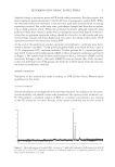
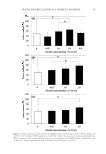
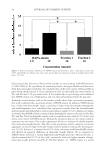
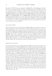
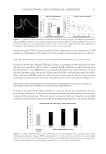
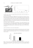
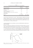
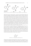
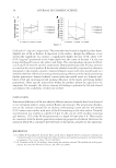
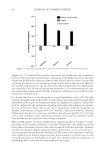
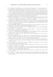
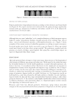
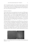
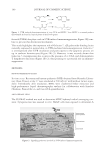
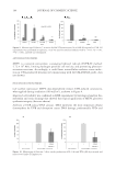
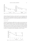
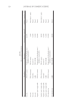
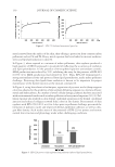
JOURNAL OF COSMETIC SCIENCE 84 Figure 6. Box chart showing expert grading of vertical ridges according to age groups. The number of data points for ages 18–30 is 75, for ages 31–50 is 105, and for ages 51–70 is 88. The severity of vertical ridges is signifi cantly higher in the age group 51–70 than the two younger groups (Tukey, p 0.001). studied here. More numerous and more profound longitudinal ridges, increased fragility and brittleness appear closely associated with aging, irrespective of ethnicity, in agree- ment with previous works (4–8) and methodologies (9). The fi ndings from the present study (as demonstrated by the clusters defi ned via PCA analysis with regard thickness) suggest that differences between fi ngernails are more driven by physical properties or their “healthy” perception. Apart from individual (or culturally/ethnically driven) desires in changing the color or shapes of fi ngernails, the development of new nail care products should then primarily aim at camoufl aging the progressive structural and physical changes of fi ngernails that are brought by an ineluc- table aging process that concerns all ethnicities. REFERENCES (1) D. Saint-Leger, The colour of the human skin: Fruitful science, unsuitable wordings, Int. J. Cosmet. Sci., 37(3), 259–265 (2015). (2) G. Loussouarn, I. Lozano, S. Panhard, C. Collaudin, C El Rawadi, and G. Genain, Diversity in human hair growth, diameter, colour and shape. An in vivo study on young adults from 24 different ethnic groups observed in the fi ve continents, Eur. J. Dermatol., 26(2), 144–154 (2016). (3) R. Baran, R. P. Dawber, and E. Haneke, Hair and nail relationship, Skinmed., 4(1), 18–23 (2005). (4) R. Baran, D. A. R. de Berker, M. Holzberg, and L. Thomas. Eds., Baran & Dawber’s Diseases of the Nails and Their Management. (Wiley Blackwell, Chichester, UK, 2012). (5) R. Baran and D. Schoon, Nail beauty, J. Cosmet. Dermatol., 3(3), 167–170 (2004). (6) G. Singh, N. S. Haneef, and A. Uday, Nail changes and disorders among the elderly, Indian J. Dermatol. Venereol. Leprol., 71(6), 386–392 (2005). (7) R. Baran, The nail in the elderly, Clin. Dermatol., 29(1), 54–60 (2011). (8) S. Murdan, Nail disorders in older people and aspects of their pharmaceutical treatment. Int. J. Pharm., 515(2), 405–411 (2016). (9) C. Ludwinski, A. Clochard-Bossuet, T. Chen, K. Norwood, and C. Oresajo, Use of optical profi lometry and visual grading for measurement of longitudinal striations of the nail, J. Cosmet. Sci., 67, 1–8 (2016).

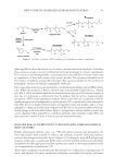
J. Cosmet. Sci., 68, 85–90 ( January/February 2017) 85 New bioprinted skin, cosmetic in vitro model SEBASTIEN CADAU, DELPHINE RIVAL, VALERIE ANDRE-FREI, MANASI CHAVAN M, DELPHINE FAYOL, MARINE SALDUCCI, BRUNO BRISSON, and FABIEN GUILLEMOT, BASF Beauty Care Solutions, Lyon, France (S.C., D.R., V.A.), Poietis, Pessac, France (D.F., B.B., F.G.), and BASF Corporation, Tarrytown, NY 10591 (M.C.). Synopsis We developed a new evolution of three-dimensional skin equivalent due to the optimization of four-dimensional laser-assisted bioprinting and skin equivalent culture protocols. This allowed us to produce fully bioprinted skin equivalents that are closed to current skin equivalents and suitable to test cosmetic ingredients. Particularly, we performed preliminary evaluation of maturogens to improve the dermis maturation before the epidermal seeding and we designed a specifi c “micropattern” to reproduce the nonlinear aspect of the dermal–epidermal junction. Finally an active ingredient was applied during the production of the bioprinted skin equivalent. INTRODUCTION Although three-dimensional (3D) printing itself is a relatively new technology, invented three decades ago, it contributes to one of the most promising medical technological advance of the century in bioscience and its market potential is only just beginning to be realized in the case of bioprinting. The fi rst description of bioprinting occurred in 1988 when R. J. Klebe described Cytoscribing, the fi rst two and 3D synthetic tissues construction on fi bronectin substrate using ink-jet printer and computer-assisted high-precision posi- tioning of cells. From the beginning of the technology development, the promises are to establish in a short timeframe precise spatial arrangements within large populations of cells to resemble natural tissues and organs for regenerative medicine or testing applications. Different printing technologies have been successfully developed using either sophisticated and complementary ink-jet, bioextrusion, or laser printers. For example, the fi rst 3D skin printing production was described in 2012 with 3D arrangement of vital cells by laser- assisted bioprinting (LaBP) as multicellular fi broblasts and keratinocytes embedded in collagen for in vitro testing application. In 2013, PrintAlive Bioprinter using complex microfl uidic device has allowed human microtissue arrays to be routinely defi ned with unprecedented speed and resolution for grafting application. Address all correspondence to Valerie Andre-Frei at valerie.andre-frei@basf.com.
Purchased for the exclusive use of nofirst nolast (unknown)
From: SCC Media Library & Resource Center (library.scconline.org)