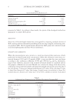
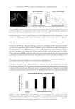
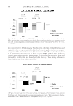
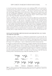
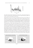
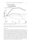
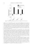
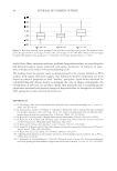
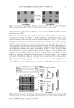
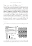
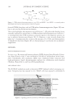
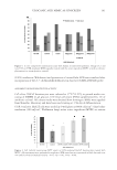
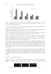
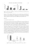
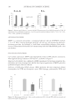
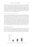
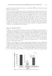
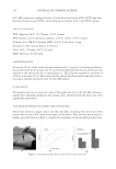
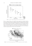
SIRT6 KNOCKDOWN 27 SIRT6 as a protective molecule in relation to the extent of DNA damage and the level of NF-κB expressed under normal conditions and after UV exposure of normal human dermal fi broblasts. MATERIALS AND METHODS TISSUE CULTURE Neonatal human dermal fi broblasts were purchased from Life Technologies. Cells were maintained in Dulbecco Modifi ed Eagle's Media (DMEM Life Technologies, Grand Island, NY) and 10% fetal bovine serum (FBS) (HyClone, GE Heathcare, Logan, UT). Cells were grown at 37ºC and 5% humidity. Donor 828840 was used at p5 and at p8 for the SIRT6 electroporation, comparing SIRT6 messenger RNA (mRNA) levels relative to RPLPO mRNA levels. Donor 871299 was used for the NF-κB experiment at passage 5. Donor 904886 was used for the fi rst comet assay, 40 mJ/cm2 UVB and 5, 10, or 20 J/m2 UVA, at passage 12. Donor 871299 was used for the second comet assay, 40 mJ/cm2 UVB and 20 J/m2 UVA, at passage 6. ELECTROPORATION OF SMALL-INTERFERING RNA The Amaxa Nucleofector II/2b Lonza Electroporation Kit (Cologne, Germany) was used to deliver small-interfering RNA (siRNA) specifi c for SIRT6. Cells were washed with Hank’s buffered saline solution before trypsinizing. An optimized electroporation pro- tocol, ID 83, from Lonza was followed. The fi broblast pellet was resuspended in the appropriate buffer and supplement (Lonza electroporation kit, VPD-1001). The Nucleo- fector II/2b was set to program U-020. Silencing was achieved using an ON-TARGETplus SMARTpool for SIRT6 from Dharmacon (Lafayette, CO) containing the following se- quences: 5′-CCAAGUGUAAGACGCAGUA-3′, 5′-GUACAUCGCUGCAGAUCCG-3′, 5′-CCAAAAGGGUGAAGGCCAA-3′, and 5′-GAACUGGCGAGGCUGGUCU-5′ or the single sequence synthesized from Dharmacon, 5′-GGAACAUGUUUGUGGAAGAUU-3′ (22). The ON-TARGETplus nontargeting (NT) pool from Dharmacon included the four following sequences: 5′-UGGUUUACAUGUCGACUAA-3′, 5′-UGGUUU ACAUGUUGUGUGA-3′, 5′-UGGUUUACAUGUUUUCUGA-3′, and 5′-UGGUUUA CAUGUUUUCCUA-3′. Each electroporation included 8–10 × 105 fi broblasts. Prior to electroporation, each cell pellet was resuspended by adding a volume of 105 μl of Lonza buffer containing siRNA at a fi nal concentration of 1 μM. The resuspended pellet was transferred to a cuvette for electroporation. Electroporated fi broblasts were seeded in four wells of a six-well plate or a 100-mm plate and incubated in 10% FBS and DMEM. RNA PURIFICATION AND REAL-TIME REVERSE TRANSCRIPTION POLYMERASE CHAIN REACTION Neonatal fi broblasts were washed with Dulbecco’s phosphate-buffered saline (DPBS) prior to extracting with 600 μl of RLT buffer from the RNeasy mini kit (Qiagen, Hilden,

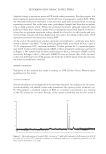
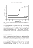
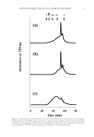
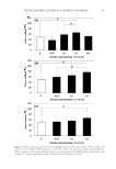
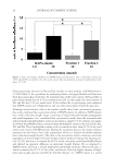
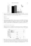
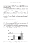
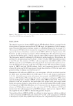
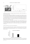
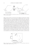
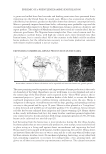
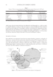
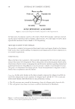
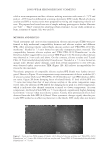
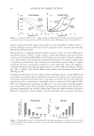
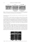
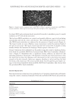
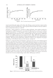
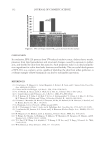
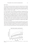
JOURNAL OF COSMETIC SCIENCE 28 Germany). The RNA extract was applied to a QIAshredder column (Qiagen) and eluted. The extract was further purifi ed according to the RNeasy protocol. The RNA concentra- tion was determined by absorbance at 260 nm with a Beckmann DU-7000 (Brea, CA) spectrophotometer (1A = 40 μg/ml). RNA was reverse transcribed with a high-capacity complementary DNA reverse transcription kit (Applied Biosystems, Foster City, CA) and amplifi ed with Taqman primer/probe sets from Applied Biosystems specifi c for human SIRT6, assay ID Hs00966001_m1, and RPLPO (60S acidic ribosomal protein P0), assay ID Hs00420895_gH. Real-time reverse transcription polymerase chain reaction (PCR) was performed and analyzed on an Applied Biosystems 7500 Fast instrument. Each PCR cycle involved 3 s at 95°C for melting and 60 s at 60°C for annealing and extension. A total of 40 cycles were completed. MODULATION OF NF-ΚB Neonatal fi broblasts were electroporated with SIRT6 siRNA and incubated for 48 h (21) before exposing the cells to 40 or 80 mJ/cm2 UVB. These doses were not cytotoxic to neonatal fi broblasts (data not shown). An irradiation chamber, BS-03 (manufactured by Dr. Grőbel UV-Elektronik GmbH, Ettlingen, Germany), was used to deliver the UVB. Six hours after irradiation, the cells were lysed and the total protein was extracted with NE-PER nuclear and cytoplasmic extraction kit from Thermo Fisher Scientifi c (Waltham, MA). Total protein was quantitated with a Micro BCA protein assay kit from Thermo Fisher Scientifi c. A chemiluminescent transcription factor detection kit from Thermo Fisher Scientifi c was used to detect NF-κB p50. A Molecular Devices Spectra- Max M2e (Sunnyvale, CA) was used to measure chemiluminescence. DETECTION OF DNA DAMAGE Each well of a Trevigen comet slide (Gaithersburg, MD) was seeded with 10,000 electro- porated fi broblasts in 200 μl of DMEM and 10% FBS. The neonatal fi broblasts were previously electroporated with SIRT6 siRNA or NT siRNA (NT). After 48 h of growth, neonatal fi broblasts were exposed to 40 mJ/cm2 UVB and 20 J/cm2 UVA using the BS-03 irradiation chamber. Cells were exposed to UV at room temperature. Cells were grown for another 4 h at 37°C and 5% humidity before processing the slides with Trevigen re- agents. No inhibitors of DNA repair were added. Slides were washed with DPBS and 75 μl of melted agarose was pipetted on each well. The cells were then processed accord- ing to the Trevigen protocol with the following changes. After 10 min at 4°C, cells were lysed for 3 h on ice. Slides were removed from the lysis solution and placed into the alka- line solution for 30 min. Slides were electrophoresed in a cold alkaline solution (300 mM NaOH, 1 mM ethylenediaminetetraacetic acid, pH 13) for 30 min at 23 V. After elec- trophoresis, slides were rinsed with water, fi xed with 70% ethanol for 5 min, and dried overnight. DNA was detected with 50 μl SYBR Green (Thermo Fisher) per well (diluted 1:10,000 in TE buffer) and incubated for 5 min at 4°C. Slides were viewed under an Olympus BX51 microscope (Center Valley, PA) using a Fluorescein Isothiocyanate fi lter and a 20× objective. Images were captured using Nikon Elements Software (Melville, NY). The tail moments were determined with Comet Score software from TriTek (Wilmington, DE).
Purchased for the exclusive use of nofirst nolast (unknown)
From: SCC Media Library & Resource Center (library.scconline.org)