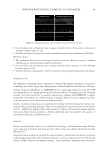
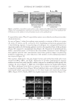
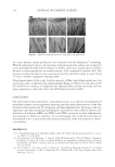
JOURNAL OF COSMETIC SCIENCE 128 the C11-fl uor probe was monitored on a fi xed number of cells (10,000) by fl ow cytometric analysis, using a FACS array system. CHELATION OF HEAVY METAL PARTICLES EPS-229, at a concentration of 0.05% w/v (to reach saturation level), was incubated for 3 h under constant agitation (200 rpm) at 25°C, in the presence of 0.3 μg/ml of Cd or Pb, in a fi nal volume of 30 ml, at pH 6. At the end of the incubation period, solutions were fi ltered by centrifugation at 3000 g, using Vivaspin 20 centrifugal fi lter units (Vivascience) with a 30 kDa molecular mass cutoff. The concentration of Cd and Pb in the supernatants was measured by fl ame atomic absorption spectrometry. PROTECTION OF SKIN EXPLANTS FROM POLLUTANT-INDUCED LIPID PEROXIDATION Skin explants were obtained from a woman (age 64) undergoing plastic surgery and maintained in culture. Every day from D0 to D4, a lotion containing the test product EPS-229 (0.03% w/v) or tocopherol (positive control) was applied at the surface of the skin explants. On D4, a small fi lter paper containing a mixture of various heavy metals plus hydrocarbons (benzene, toluene, xylene, anthracene, and naphthol) was additionally applied at the surface of the skin explants. On D5, fi lter papers were removed and malondialdehyde (MDA) levels, the end product of lipid peroxidation, were quantifi ed in the culture media of each skin explant, using enzyme-linked immunosorbent assay. PROTECTION OF SKIN EXPLANTS FROM POLLUTANT-INDUCED MORPHOLOGICAL CHANGES On D5, at the end of the pollutant challenge experiment described earlier, skin explants were treated with Bouin’s histological reagent for 48 h, dehydrated, and paraffi nized. Morphological studies were done on cut paraffi n sections, following Masson’s trichrome staining. CLINICAL EVALUATION The effi cacy of EPS-229 to protect the skin against air pollutants was evaluated on a panel of 18 healthy women, aged 42–72 years. Microparticles of black iron oxide with a size of 1 μm were used, as a mimic of the PM (PM2.5) released in the atmosphere by industrial activity and motor vehicle emissions (6). For the anti-adhesion study (preexposure action), two zones (A and B) were defi ned on the forearms of volunteers. Zone A was treated with EPS-229 (0.02% w/v) in a water solution and zone B with a water placebo for 20 min. Next, a solution containing black iron oxide microparticles was applied on both zones using a make-up sponge. Three minutes later, both zones were rinsed with water (4 μl/cm2) and then wiped to remove nonadherent particles. Pictures of both zones were taken before and after rinsing, with a Hirox® video microscope. The percentage of nonadherent PM2.5 particles was calculated using the fol- lowing formula: ((number of adherent PM2.5 particles before rinsing - number of adherent PM2.5 particles after rinsing)/number of adherent PM2.5 particles before rinsing) × 100.

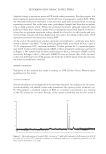
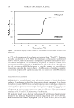
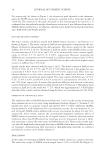
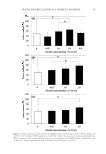
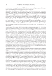
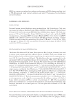
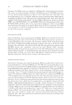
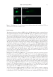
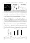
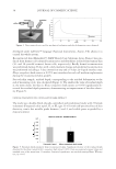
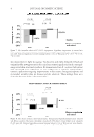
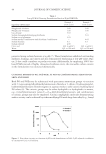
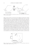
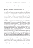
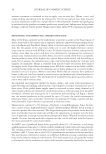
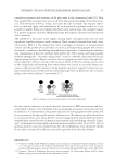
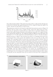
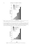
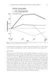
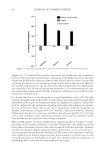
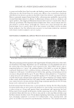
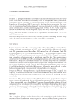
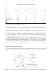
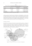
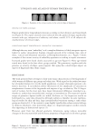
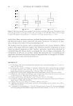
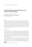
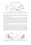
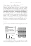
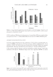
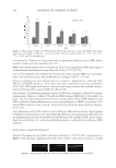
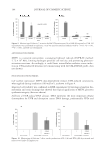
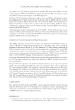
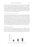
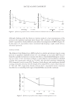
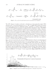
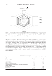
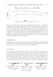
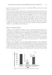
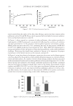
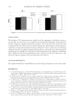
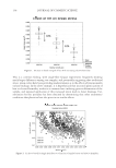
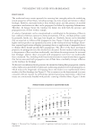
EXOPOLYSACCHARIDES FOR BIOMIMETIC PROTECTIVE EFFECT 129 Statistical analysis was done using normality test, Shapiro–Wilk test, Student’s t test and Wilcoxon signed-rank test. For the cleansing study (postexposure action), the protocol was modifi ed as follows: water (2 μl/cm²) was fi rst applied on zone A and zone B, and skin left to dry. A solution contain- ing black iron oxide microparticles was next dropped on both zones using a make-up sponge. Three minutes later, zone A was rinsed with EPS-229 (0.01% w/v) in water solu- tion and zone B with water, massage, and wiped with a dry cotton tissue to remove non- adherent particles. Pictures of both zones were taken before and after rinsing, with a Hirox® video microscope. The percentage of removed PM2.5 particles was calculated us- ing the following formula: ((number of adherent PM2.5 particles before cleansing – number of adherent PM2.5 particles after cleansing)/number of adherent PM2.5 particles before cleansing) × 100. Statistical analysis was done using normality test, Shapiro–Wilk test, Student’s t test and Wilcoxon signed-rank test. RESULTS AND DISCUSSION In Figure 1, results obtained in fl uorescence have been transformed to express protection of keratinocyte membranes from radical formation. Not surprisingly, UV exposure of cells resulted in a signifi cant increase (expressed as 100% from baseline) in the level of lipid peroxidation at the membrane of cells. Addition of the synthetic antioxidant BHA (positive control) reduced the formation of UV-induced lipid peroxidation by 76%, vali- dating the assay. In the presence of EPS-229, protection reached 28% for a concentration of 0.001% w/v. Thus EPS-229 is able to effectively protect skin cells from UV-induced lipid peroxidation. Protection from UV-induced lipid peroxidation is associated with prevention of skin photo-aging (7). In Figure 2, data obtained in tubo from the heavy metal adsorption studies confi rmed the chelation potential of EPS-229 toward cadmium and lead. In both cases, metallic retention reached a plateau allowing for the calculation of the maximal adsorption (Qmax) potential of EPS-229 for these cations, in accordance with the Langmuir adsorption model. For Cd, Qmax was estimated at 154 mg/g (1.37 mmol/g). For Pb, Qmax was estimated at 250 mg/g (1.21 mmol/g). The ability of EPS-229 to adsorb divalent cations may facilitate heavy Figure 1. EPS-229 protects keratinocytes from lipid peroxidation.
Purchased for the exclusive use of nofirst nolast (unknown)
From: SCC Media Library & Resource Center (library.scconline.org)