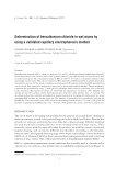
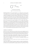
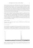
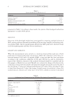
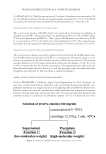
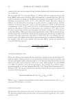
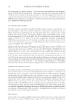
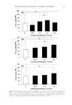
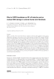
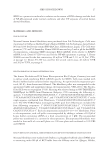
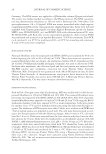
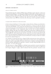
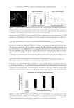
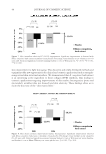
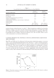
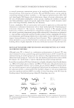
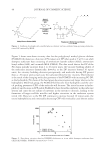
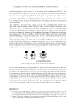
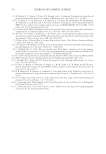
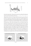
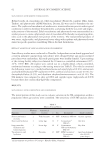
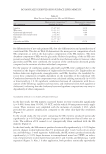
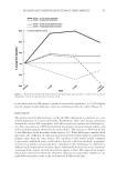
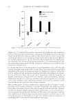
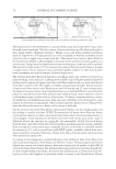
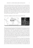
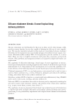
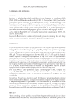
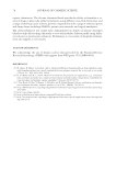
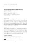
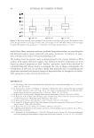
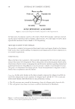
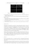
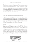
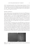
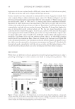
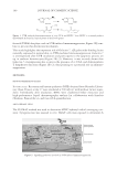
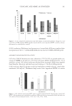
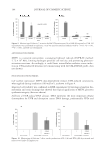
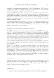
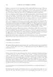
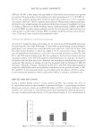
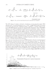
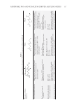
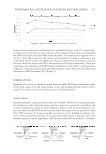
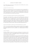
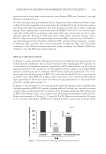
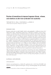
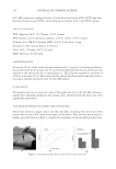
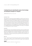
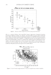
SIRT6 KNOCKDOWN 29 RESULTS SIRT6 KNOCKDOWN Neonatal human dermal fi broblasts were electroporated with SIRT6 siRNA and the mRNA levels for SIRT6 were monitored 48 h after electroporation. A 72% decrease in SIRT6 mRNA was achieved with four siRNAs and a 68% decrease with one SIRT6 siRNA (Figure 1). The NT siRNA level of SIRT6 mRNA was comparable to the untreated. IMPACT OF SIRT6 KNOCKDOWN ON NF-κB SIRT6 knockdown elevates NF-κB p50 levels signifi cantly in the absence of UVB (Figure 2). NF-κB p50 levels were monitored 52 h after electroporation using an enzyme-linked Figure 1. Neonatal fi broblasts were electroporated using four siRNAs specifi c for SIRT6 or a single siRNA for SIRT6 (22). An average of 70% knockdown was observed after 48 h relative to the nontargeted cells from three separate experiments. Data are expressed as mean ± SEM, *p 0.005. Figure 2. Neonatal fi broblasts were electroporated with SIRT6 siRNA and incubated for 48 h before expos- ing to UVB. Six hours later, cells were lysed. The nuclear fraction (squares) was separated from the cytosolic faction (diamonds) and probed for NF-κB p50 (NT-cy, -nu = non-targeting cytosolic or nuclear SIRT6 KD-cy, -nu = SIRT6 knockdown cytosolic or nuclear). In the absence of UVB, SIRT6 knockdown increased NF-κB translocation to the nucleus fourfold relative to NT. Data expressed as mean ± SEM, *p 0.05.

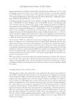
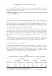
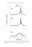
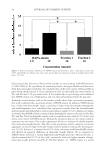
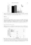
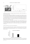
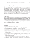
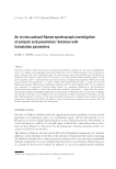
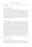
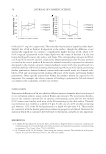
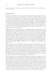
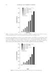
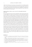
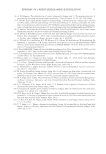
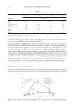
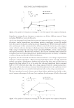
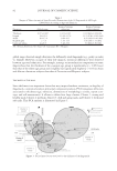
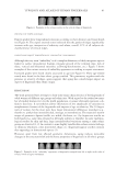
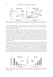
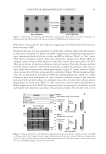
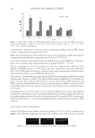
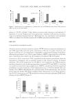
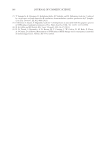
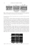
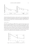
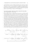
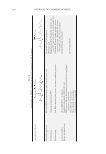
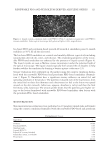
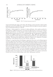
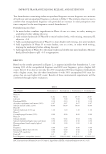
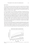
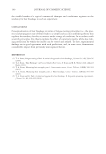
JOURNAL OF COSMETIC SCIENCE 30 immunosorbent assay antibody detection assay. A 4.3-fold increase in chemiluminescence was observed in cells receiving SIRT6 siRNA relative to NT siRNA. A transient reduc- tion in SIRT6 mRNA levels was suffi cient to elevate NF-κB p50 levels more than four- fold relative to the NT cells. In the presence of UVB, the SIRT6 knockdown fi broblasts continued to generate more chemiluminescence relative to the NT fi broblasts, but the differential decreased as the UV dose increased (Figure 2). Exposure of the SIRT6 knockdown cells to 40 or 80 mJ/cm2 of UVB increased NF-κB p50 levels about fi vefold and sixfold, respectively, to SIRT6 knockdown cells not receiving UVB. However, the differential between the SIRT6 knockdown cells and the NT cells after receiving 40 mJ/cm2 was about 1.5-fold and after receiving 80 mJ/cm2, the differential was 20%. Cytosolic levels of NF-κB p50 remained low in the presence or absence of UVB. Thus, the largest differential increase in NF-κB levels of the SIRT6 knockdown cells versus the NT fi broblasts oc- curred without UV exposure. The addition of UV increased nuclear NF-κB levels for both the NT and the SIRT6 knockdown, but the differential decreased as the dose of UV increased. IMPACT OF SIRT6 KNOCKDOWN ON DNA DAMAGE In the absence of UV, SIRT6 knockdown increases the tail moment about 10-fold relative to the NT fi broblasts (Figure 3). Cells were lysed 4 h after UV exposure. Examination of nuclear DNA by fl uorescent microscopy with SYBR Green staining revealed a circular shape for the NT fi broblasts and the SIRT6 KD cells, refl ecting a compact nucleus (Figure 4). In the absence of UV, SIRT6 KD cells had a visible tail. After UV, SIRT6 knockdown cells increased their tail moment about twofold relative to the NT fi broblasts (Figure 3). Cells were allowed 4 h to repair the DNA damage resulting from 40 mJ/cm2 UVB and 20 J/cm2 UVA. The nucleus appeared fractured for the NT fi broblasts and the SIRT6 KD cells with a visible comet tail (Figure 4). Figure 3. Neonatal fi broblasts seeded on comet slides were exposed to 40 mJ/cm2 UVB and 20 J/cm2 UVA. Cells were grown for another 4 h before processing the slides. SIRT6 knockdown increased the tail moment about fi vefold relative to the nontargeted, whereas no signifi cant change was observed when UV was added. Data are expressed as mean ± SEM, *p 0.05.
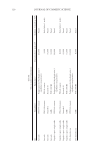
Purchased for the exclusive use of nofirst nolast (unknown)
From: SCC Media Library & Resource Center (library.scconline.org)