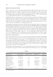
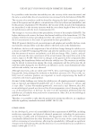
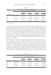
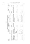
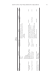
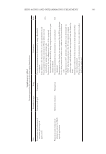
Title Formulation type Drug Evaluation Refs. In vitro and in vivo skin antiaging evaluation of gel containing niosomes loaded with a semi- purifi ed fraction containing gallic acid from Terminalia chebula galls Gel containing niosomes Semi-purifi ed fraction containing gallic acid In vitro biological antioxidant activity of gallic acid and the semi purifi ed fraction incorporated into the optimized elastic and non-elastic niosomes was estimated via the free-radical scavenging (96) Cytotoxicity assay was performed via MTTjassay MMP-2kinhibition activity by gelatinolytic zymography (gelatinolytic activities of MMP-2 was assessed by SDS-PAGEl zymography using gelatin as a substrate) Evaluating the physicochemical stability of the gel containing niosomes loaded with the semi-purifi ed fraction was carried out Skin irritation tests on male rabbits (irritation index depends on erythema and edema degree) Effi cacy investigation of the antiaging potential of the optimized gel in human volunteers was detected via determination of skin elasticity, surface microstructure, hydration, erythema and pigmentation Development and evaluation of vesicular system for curcumin delivery Phyto-vesicles, liposomes and niosomes Curcumin Characterization of curcumin–phosphatidyl choline complex was performed via TLCm, DSCn, melting point, and FTIRo (97) Characterization of the vesicular systems Morphological study by TEM Vesicles size and PDIp by Malvern Zetasizer Spectrophotometric determination of the entrapment effi ciency Assessment of the anti–aging capability of the developed vesicular systems in UV-radiated Swiss albino mice was carried out by examining certain biochemical markers, moisture content, and histological analysis Effects of skin penetration enhancers in topical antiaging products containing α-hydroxyacids and hyaluronic acid Conventional lotion α-Hydroxyacids Evaluating the effect of different permeation enhancers on transdermal penetration was performed by using skin permeation tests via diffusion cells (72) Hyaluronic acid Supplementary table I Continued JOURNAL OF COSMETIC SCIENCE 346

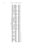
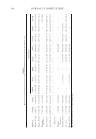
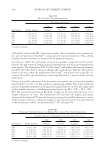
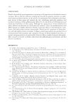
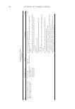
Title Formulation type Drug Evaluation Refs. Transdermal delivery of functional collagen via polyvinylpyrrolidone microneedles Polyvinylpyrrolidone microneedles Collagen type 1 Examination of the microneedle morphology was detected via SEM and CLSMq. (77) Collagen type 1 was separated via SDS-PAGE electrophoresis Functional collagen concentration was determined via ELISAr Microneedle penetration through the porcine skin and human foreskin was evaluated via a fl uorescence microscope Transdermal delivery of proteins mediated by non-covalently associated arginine-rich intracellular delivery peptides Arginine-rich intracellular delivery (AID) peptides Protein Transdermal delivery assay on mice (histological examination) (83) Protein internalization on human A549 cells(confocal s microscope) Cytotoxicity in Human A549 cells (MTT assay) A peptide carrier for the delivery of elastin into fi broblast cells Amphipathic cell– penetrating peptide carrier Elastin The SDS-PAGE technique was used to determine protein/peptide complex formation (85) Particle size distribution by DLS Particles morphology was determined via SEM. Cytotoxicity study in NIH-3T3tcells (MTT assay) Internalization of the complex in NIH-3T3 cells was evaluated using a fl uorescent microscope CoQ10enhances dermal elastin expression, inhibits IL-1α production and melanin synthesis in vitro O/W Nano emulsion CoQ10(CoQ 10 ) Investigation of CoQ10anti-aging effect in multiple adult fi broblast cell lines was performed by using a cell proliferation assay. (99) Illustrating the effect of CoQ10on ROSproduction u by radiating fi broblast cell lines with UV radiation followed by measuring the intracellular ROS level Evaluating the depigmentation potential of CoQ10was detected via melanin assay, tyrosinase activity measurement and DOPAvstaining Systematically optimized CoQ10- loaded proniosomal formulation for treatment of photo-induced aging in mice: characterization, biocompatibility studies, biochemical estimations and anti-aging evaluation Proniosomal gel formulation CoQ10 In vitro characterization of the proniosomal gel: (100) The % drug entrapped was determined by using HPLC method Particle size analysis was measured by using a Malvern zetasizer Morphological studies were detected by using TEM Rheological studies were determined by using a cup-and-bob viscometer. Ex vivo evaluation of the drug permeation and retention was performed. In vivo evaluation of the drug antiaging effect in female Swiss albino mice after exposure to UV radiation was carried out via visual, histopathological and other evaluations Supplementary table I Continued SKIN-AGING AND INFLAMMAGING TREATMENT 347
Purchased for the exclusive use of nofirst nolast (unknown)
From: SCC Media Library & Resource Center (library.scconline.org)