JOURNAL OF COSMETIC SCIENCE 240 construction (1). The fruits are large (20 cm), with hard shell and a cover that opens when mature, releasing light brown seeds. The fruits form from June to September. The seeds germinate in 40–70 d, are edible, are tasty, and possesses medicinal proprieties (1). The olea ginous seeds of L. pisonis are rich in proteins, lipids, fi ber, thiamine, ribofl avin, niacin, phosphorus, and potassium (3–5) and reasonable amounts of calcium and magne- sium (4,5). Their low sodium content and no cholesterol is benefi cial for the cardiovascular system (3,5,6). Therefore, they are considered a functional and nutritional food for humans (6). In tradi tional medicine, L. pisonis leaves are used to treat pruritus and muscle pain, their analgesic effect in humans might be related to their antinociceptive effects in mice (6). The anti oxidant activity of the ethanolic extract of L. pisonis leaves may be associated with high levels of polyphenols and fl avonoids (7). Oil from L. pisonis showed antioxidant activity that may be related to tocopherols, α and β-tocopherol, and vitamin E (4). The aim of this work was to develop a skin cream using L. pisonis nut oil and evaluate its quality to add value to the oil as a raw material and to the plant as a living resource. This will reduce the need to raze L. pisonis trees and promote the maintenance of this species and sustainable development in the Amazon region, creating a potential source of revenue for the region. MATERIALS AND METHODS PLANT MATE RIAL Fruits of L. pisonis were harvested in Laranja da Terra, Espírito Santo, Brazil, as a part of the 2015 crop acquired by the Instituto Capixaba de Pesquisa e Extensão Rural. The voucher specimen was deposited in the Coleção de Herbário do Jardim Botânico do Rio de Janeiro (JBRJ-Holotype) and Royal Botanic Gardens (K000600113). After selection, where bruised nuts where discarded, the nuts were divided into four groups and stored at –18°C until analysis. The nuts were peeled and macerated with hexane in a Soxhlet (Unividros, Ribeirão Preto, Brazil) apparatus for 6 h to obtain the oil according to the International Union of Pure and Applied Chemistry method 1.122 (8). After the solvent was removed by evaporation under reduced pressure, the oil was stored in an amber glass bottle under refrigeration (–8°C) until use. PHYSICAL–CHEMICAL ANALYSIS OF THE OIL OF L. PISONIS NUTS Acidity index of t he oil was determined according to the physical–chemical methods for food analysis (9), with modifi cations. Briefl y, 2 g of the sample was dissolved in 25 mL of a 2:1 ether:alcohol mixture and was titrated with a standard solution of 0.01 M potassium hydroxide (KOH) using phenolphthalein as an indicator. The acidity index was calcu- lated and expressed in mg KOH/g oil. The peroxide index was also determined according to the physical–chemical methods for food analysis (9), with modifi cations. The oil (5 g) was shaken for solubilization with 30 mL of an (3:2) acetic acid:chloroform mixture. Then, protected from light, 0.5 mL of saturated

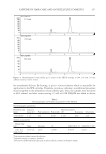
USE OF LECYTHIS PISONIS OIL IN COSMETIC CREAMS 241 potassium iodide solution and 30 ml of water were added. Thereafter, the mixture became yellow and was titrated with a standard solution of 0.1 N sodium thiosulfate until the yellowish color was not perceptible anymore. At this point, on adding 0.5 mL of starch indicator solution, the solution turned blue and titration with a standard solution of 0.1 N sodium thiosulfate was continued until the blue color disappeared. The peroxide index was calculated and expressed as meq KOH/100 g sample. Also, the lipids a nd fatty acids in the oil and formulations were determined, where the fat was extracted from the samples according to the Bligh–Dyer method (10). Briefl y, 2.5 g of the sample was mixed with a (10:20:8) solution of chloroform, methanol, and milli-Q water. The mixture was shaken and centrifuged at 900 rpm for 30 min, and then 10 mL of chloroform and 10 mL of aqueous solution of 1.5% sodium sulfate were added and shook again for 2 min. Then, the chloroform phase containing the fat was collected and fi ltered. After the removal of the solvent by evaporation with under-reduced pressure, the total fat content was determined by gravimetry. The fatty acids ex tracted from the sample were converted to fatty acid methyl esters (FAMEs) according to Joseph & Ackman (11), using 10% BF3 in methanol. Briefl y, 15 mg of the extracted fat was added to 1.5 mL of 0.5 M sodium hydroxide in methanol in a capped tube. The closed tubes were placed in a boiling water bath for 5 min. After cooling at room temperature, 2 mL of 10% BF3 in methanol was added, and the tubes were returned to boiling for 30 min. After another cooling step, 1 mL of isooctane was added and shook for 30 s. Finally, 5 mL of saturated sodium chloride solution was added to the tubes and shaken. The isooctane phase (containing FAMEs) was collected. As control, 1 mg·mL-1 methyl trioctanoate (C23:0Me) was added to the samples, to allow the correction of FAME quan- tifi cation due to variability in extraction, analytical instrument, or solvent evaporation. The resulting FAMEs w ere injected into a Shimadzu GC-2014 gas chromatograph (GC) with a fl ame ionization detector (FID) and an HP-INNOWax (Agilent, Santa Clara, CA) capillary column (50 m × 0.20 mm i.d. × 0.20 μm). The chromatographic conditions were as follows. The injector was operated at 250°C in split mode (1:10) for 1.0 min. The nitrogen drag gas fl ow was 1.25 mL/min, and the detector temperature was 260°C. The oven temperature gradient program was as follows: an initial temperature of 150°C, which was increased at 10°C/min to 260°C, where it was held for 9 minutes. A standard solution of FAMEs (GLC-85, Nu-check) was injected into the GC-FID system under the same conditions as for the samples. All analyses were performed in triplicate. The fatty acids were quantifi e d according to Visentainer (12) using the FAME areas in the chromatograms the areas were corrected with the theoretical correction factors (TCFs) and the conversion factors for fatty acids to FAME obtained using internal standards. To quantify the metals contain ed in the oil, nuts, arils, shells, and oil were digested with 10% nitric acid (65% purity, Sigma-Aldrich, Darmstadt, Germany) at 150°C using a Marconi digester (model MA 851, Marconi Equipamentos para Laboratório LTDA, Piracicaba, Brazil) and fi ltered through a 0.22-μm fi lter (Jet Biofi l, Guangzhou, China). Flame atomic absorption spectrophot ometry quantifi cation was performed in triplicate, using an atomic absorption spectrophotometer (iCE 3000, AA05141602 v.1.30, Thermo Scientifi c, Waltham, MA) and an atomizer with an air/acetylene burner and a hollow cathode lamp (Photron PTY. Ltd., Narre Warren, Australia) as a source of radiation to determine target elements. The calibration curve ranges for each analyte were as follows: Fe (1–15 mg/L), Na (1–15 mg/L), and Pb (10–40 mg/L). The chemicals were analytical
Purchased for the exclusive use of nofirst nolast (unknown)
From: SCC Media Library & Resource Center (library.scconline.org)