712 JOURNAL OF COSMETIC SCIENCE thinner diameters tend to have tighter curl. It has also been shown that the lipid content in African hair is greater than in other hair types (8–10). Developing an understanding of hair curvature has challenged scientists for some time and has led to a significant amount of work in this area (11–20). A number of hypotheses have been proposed to explain hair curvature, and they involve follicle anatomy, asymmetric expression of structural keratins in the precortex, variable cortical shape, keratin filament orientation (relative to the hair growth axis), asymmetric proliferation in cells that form the inner and outer root sheaths, and dermal papilla asymmetry (21). Examining single fibers, the intrinsic optical properties (luster) of hair obtained from various ethnic origins were determined by researchers at TRI Princeton (22). They found that African and Indian hair had the greatest luster however, it should be noted that the degree of hair color greatly influences the outcome of such results. Also, they carried out goniophotometer measurements of single stretched fibers. Luster measurements carried out on hair in its natural state are more realistic and reflect the complexity of the hair fiber assembly and its contributions to the overall optical properties, especially in textured hair (23). Interestingly, the amino acid composition is the same for all hair types, which has been reported in several studies (24,25). A proteomics study of South Africans of indigenous African, Indian, Caucasian, and mixed ancestry was completed and identified several different protein classes (keratins, keratin-associated proteins, histone proteins, and desmosomes) in hair. However, they were unable to find a quantitative distinction between the different proteins found in each group of subjects (26). But a genome-wide association test of people living in Latin America with mixed European, Native American, and African ancestry reported a substitution on the PRSS53 protein from the protease serine S1 family that is associated with scalp hair curvature (27). As the traditional continental boundaries of race diminish, proteomics and genomics studies are becoming more important in identifying hair traits associated with racial ancestry. One of the principal motivations for understanding the behavior and physicochemical properties of textured hair stems from unique cosmetic treatment protocols that are used by populations with these hair types. For example, hair straightening and relaxing remain popular hair procedures to modify the overall 3D structure of hair fiber assemblies. Historically, interest in elucidating structural changes to the hair fiber associated with these treatments led to a better understanding of overall hair damage (28–32). MATERIALS AND METHODS Studies were carried out on dark brown European (Caucasian) and two types of African hair purchased from International Hair Importers and Products Inc. (Glendale, NY) and DeMeo Brothers, Inc. (Passaic, NJ). The European hair and one type of African hair (referred to as tightly curled African hair throughout the text) were obtained from International Hair Importers and Products Inc. We purchased another type of African hair (referred to as extremely tightly curled African hair) from DeMeo Brothers, Inc. For the analyses, many fibers were collected from a single hair tress for each hair type. While the Caucasian hair was blended, the African hair types came from one individual. Before analysis, all hair was washed with a 3% (w/w) sodium laureth sulfate:cocamidopropyl betaine (12:2) mixture.

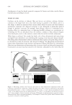
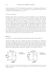
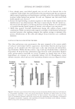
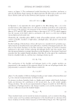
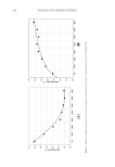
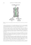
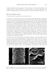
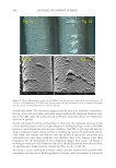
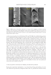
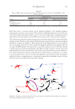
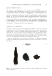
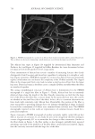
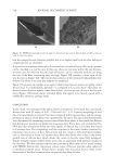
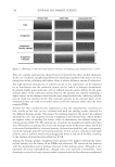
713 PHYSICOCHEMICAL PROPERTIES OF TEXTURED HAIR MACROPHOTOGRAPHY OF HAIR Photographic images of hair were obtained using an Epson Perfection V550 Photo Color Scanner (Epson Seiko, Suwa, Japan) with Epson Scan (version 3.9.1.1US) software. Images were collected in 24-bit color mode at 300 dpi. Image analysis of the photographs was carried out with ImageJ (version 1.53a) software (National Institutes of Health, Bethesda, MD). The lengths of individual hair fibers in the relaxed and fully extended states were measured to determine the curl index (CI) of hair. HAIR CROSS-SECTION PREPARATION Hair obtained from tresses was sectioned using a Leica CM3050 S (Leica Microsystems GmbH, Wetzlar, Germany) cryostat equipped with a high-profile sectioning head and Leica 818 high-profile cutting blades. Note that the cryostat blade was changed after each sample. For each sample, a 25 mm aluminum specimen disk was pre-equilibrated in dry ice (−78.5°C). A small section (1/8–1/4 in. wide × 1–2 in. long) of the ¾ in. wide tress was removed from the center of the tress length. The tip-end of the damp fiber section was then held straight and perpendicular against the platform of the disk. At the interface between the disk and tip-end of the fiber bundle, a drop of distilled water was then added. The drop instantly crystallized, hence causing the tip-end of the fiber bundle to adhere to the aluminum specimen disk. By pulling gently upward on the root end of the fibers and slowly adding water to the fiber bundle, a straight rod of ice-embedded hair was produced. Separately, each embedded sample was then mounted onto the specimen head (−30°C) of the cryostat, where the chamber temperature was equilibrated at −25°C. After conditioning for at least 1 h, 5-μm–thick sections were collected in continuous mode using a sectioning speed of 60% maximum. Hair cross-sections were air-dried on paper overnight under reduced pressure. FIELD EMISSION SCANNING ELECTRON MICROSCOPY (FESEM) OF HAIR The cross-sections were imaged using field emission scanning electron microscopy (FESEM) to investigate the internal morphological structure of hair and to calculate the cortical cross-sectional area (n=100). Cross-sections were fixed to aluminum Pelco pin stubs using 25 mm conductive carbon tabs (Ted Pella, Redding, CA) and then coated with Au/Pd using our Leica EM ACE600 sputter coater. Finally, the hair fiber cross-sections were imaged with a Hitachi SU-5000 FESEM (Hitachi High Technologies, Schaumburg, IL) using various magnifications. FOURIER TRANSFORM INFRARED (FTIR) IMAGING OF HAIR CROSS-SECTIONS Fourier transform infrared (FTIR) images were obtained with a PerkinElmer Spotlight 400 FTIR imaging microscope (PerkinElmer Inc., Waltham, MA), which combines an optical microscope with an FTIR spectrometer. The system consists of a linear array of mercury- cadmium-telluride detectors coupled to a precision automated X–Y sampling stage. Background spectra were collected on sample-free areas of the CaF 2 crystal, and FTIR images were obtained at 8 cm−1 spectral resolution in transmittance mode at 16 scans/pixel.
Purchased for the exclusive use of nofirst nolast (unknown)
From: SCC Media Library & Resource Center (library.scconline.org)