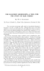
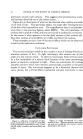
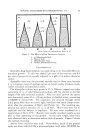
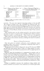
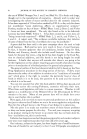
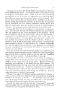
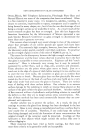
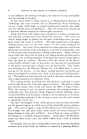
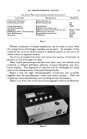
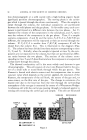
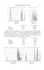
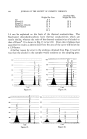
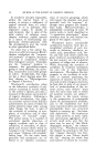
THE ELECTRON MICROSCOPE--A TOOL FOR THE STUDY OF HAIR FIBERS* By W. L. COURCHENE The Procter & Gamble Co., Miami 1/alley Laboratories, Cincinnati $7, Ohio THE ELECTRON microscope really needs no introduction because it has been in use for a number of years. During these years it has been applied to a wide variety of problems, but the application of electron micros- copy to hair and related problems has received little attention (1, 2, 3). It is the main purpose of this paper to point out to those interested in hair and hair products the potential usefulness of certain techniques of electron microscopy. The electron microscope makes available certain magnifications that could not previously be obtained by other methods. In terms of resolving power, it bridges the rather large gap between the magnifications available with light microscopy and those available with x-rays. Figure 1 shows this in a better way. This drawing has been constructed to show the structural details of a single hair fiber by the device of increasing the magnification from left to right. For those who are not familiar with the hair structure it consists of a thin layer of cells on the outside called the cuticle, and a central cortex composed of spindle-shaped cortical cells. These are 7-103t wide and 1003t long. Within these cortical cells one finds macrofibrils. At still higher levels of magnification one notes that these macrofibrils are composed of still smaller fibrils called microfibrils. The light microscope can be used only to a magnification of about a thousand times, so that the •uticle, cortex and sometimes cortical cells can be distinguished, along with scale patterns on the surface. X-rays, on the other hand, can tell us some- thing about atomic and molecular arrangements at a magnification of about a million (106). X-rays do not give a picture as the electron microscope does, but information can be derived from the patterns. The development of the electron microscope has made possible the observation of the histological structures intermediate between cortical cells and molecules by making available magnifications between 1000 and 100,- * Presented at the December 13, 1956, Meeting, New York City. 60

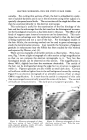
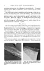
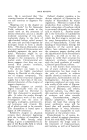
ELECTRON MICROSCOPE--TOOL FOR STUDY OF HAIR FIBERS 61 0 I Magnification 000. Even in its lower range of magnification, where electron microscopy overlaps light, the electron microscope brings out details which cannot be clearly observed with light microscopy. That is, the usefulness of the electron microscope for hair is not confined to the examination of structures in the range shown in Fig. 1, but can be used to greatly magnify those features of the hair which are ordinarily visible in a light microscope. Since it is now possible to examine the fine structure of hair, I would like to discuss several techniques which can be used, the kinds of problems to which these techniques can be applied, and show some examples of the kind of electron micrographs obtained. Of the various techniques which have been developed for use in electron microscopy, we believe three of these basic techniques of sample preparation to be the most useful for the study of hair. These are the replication tech- nique for examining surfaces, the ultrathin sectioning technique for examin- ing interior histological structures in situ, and the degradation technique for looking at specific histological structures as individual particles separated from the other structures. REPLICATION Of the three, the replication technique is probably the simplest to use. A i thin, electron-transparent cast is made of the hair surface and examined. IThere are many replication techniques in existence today which have been I developed to examine specific kinds of surfaces. Of these techniques, •we believe the one using evaporated metal to be generally the most satis- I 0 I0 z 10 3 10 4 10 5 10 8 I I I I I I Cortical Macro Micro Cells Fibrils Fibrils Atoms Fiber Cuticle Molecules I•X-raY s I Electron Microscope---I I Light Microscope I Figure 1,--Diagram of a single hair fiber progressively magnified to show the histological I structure. Magnifications are only approximate. The useful range of the light and electron microscopes and of x-rays is shown.
Purchased for the exclusive use of nofirst nolast (unknown)
From: SCC Media Library & Resource Center (library.scconline.org)