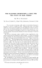
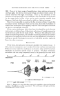
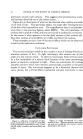
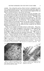
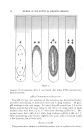
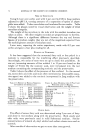
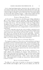
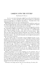
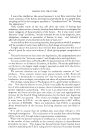
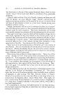
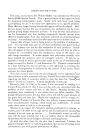
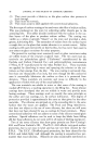
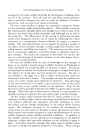
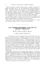
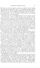
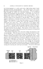
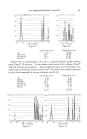
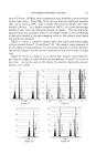
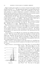
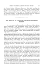
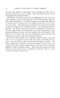
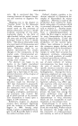
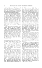
JOURNAL OF THE SOCIETY OF COSMETIC CHEMISTS previously smooth scale surfaces. This suggests that proteinaceous mate-• rial has been dissolved out of the surface layers. Figure 6 is an illustration of what can be observed when replicas are made• of cut ends of hair. This particular replica was made after etching the end I of the hair with hydrochloric acid. The magnification here is considerablI more than in the previous figures, about 8000X. The cell membrane ofl cortical cells is plainly visible, and one cortical cell is outlined in its entirety.: In the center is what appears to be the dead nucleus of this cortical cell. I Also faint outlines of macrofibrils are visible just below the nucleus. These examples are just a few of the many effects which can be observed in hair by this replication technique. ULTRATHIN SECTIONING The second technique useful for hair studies is that of looking directly at• ultrathin sections of hair. In light microscopy thin sections are of the l order of a few microns thick. In electron microscopy these sections must• be a few hundredths of a micron thick because of the lesser penetratingl power of electrons compared to light. There are microtomes for cutting I ultrathin sections which have become commercially available in the past• couple of years. Our microtome happens to be a Sjostrand thermal ad-I vance device, but a Porter-Blum mechanical advance microtome is also• Figure 6.--Electron micrograph of chro- mium metal replica of an end of a hair fiber exched with dilute hydrochloric acid. Corti- cal cell walls, a nudeus and macrofibrils are visible. Figure 7.--Light micrograph of a cross section of hair. Pigment granules and corti-i cal walls are visible but no structure can be• discerned in the cuticle.

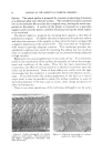
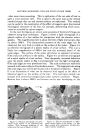
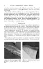
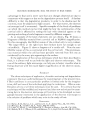
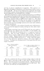
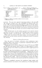
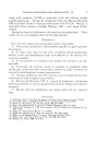
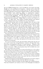
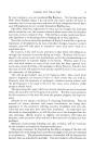
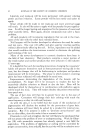
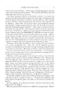
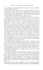
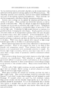
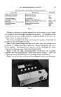
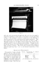
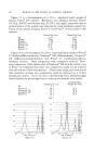
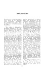
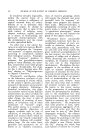
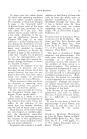
ELECTRON MICROSCOPE--TOOL FOR STUDY OF HAIR FIBERS 65 suitable. For cutting thin sections of hair, the hair is embedded in a poly- mer of suitable hardness and is cut in the microtome using either a glass or a specially sharpened metal knife. The cut sections of the single hair fiber can then be examined directly in the electron microscope. This technique is useful for the examination of the fine histology of the hair and has the advantage that the hair need not be disintegrated to exam- ine the histological structures as has been done in the past. The effect of all kinds of reagents upon internal structures can be determined. This tech- nique has an advantage over the replication method in that the hair itself is being examined and not a cast of the hair. For histological studies in the hair follicle, the sectioning method can be used to great advantage to study the keratinization process. Just recently the formation of pigment granules in melanocytes near the follicle has been studied by this method and reported by Mercer, et al. (4). There are two examples of ultrathin sections as illustration. First, how- ever, Fig. 7 shows a light micrograph of a thin section at high magnification so that we can compare the electron micrographs to it. Very few fine histological details can be observed on this section. The magnification is about 760X, slightly less than the maximum obtainable. The cuticle of the hair can be distinguished along the edge but no structure is apparent within it. Pigment granules are also visible as small dark dots. Cortical cell walls are visible as lighter lines in the red background caused by staining. Figure 8 is an electron micrograph of an ultrathin section of hair at about 4300) magnification. It is seen that the cuticle is composed of thin cells that are arranged concentrically around the cortex of the hair. The cortical cell walls are very prominent. The dark spots are pigment granules. Sev- "?' ..lj !"*½ • "' '.:. y •*:-- •,. . ..... • . . ' .. ... 3 •- . ::7.: '.'.. -:• '• .::-•"• T" •:' --' -•- '*•: •' ......... :} ::..'.'•:.... ß :....: Figure 8.•Electron micrograph of ultra- thin cross section of hair. Cortical cell walls, pigment granules, cuticle cell walls, and cortical cell nuclei are visible. •t' '•'•'C.'."" . •:w" .•- ,• •. :t•'. .:.?... -'•: ,... :• o. t:.. • ..:• -•- 4. .... :•! •.. ..• '.•:.ii'"' ,. •--•'i . .:.• • - % ... :. ..... : . .... :. ,• •.: . -.• . c . •. -- - :• ..:.• • .... * ... : : '*. ,-,- : .... ... ... , ' ... . ...... .. ..m .. •... Figure 9.•Electron micrograph of ultra- thin cross section of hair. Note the over- lapping of cuticle cells.
Purchased for the exclusive use of nofirst nolast (unknown)
From: SCC Media Library & Resource Center (library.scconline.org)