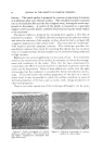
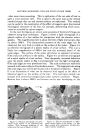
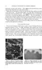
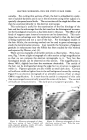
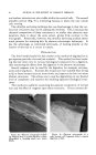
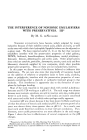
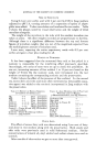
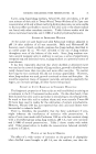
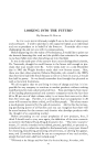
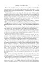
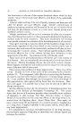
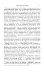
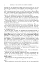
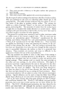
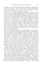
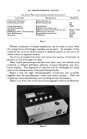
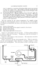
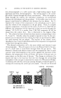
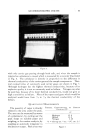
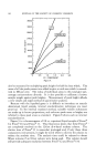
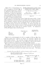
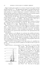
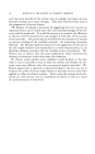
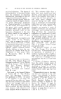
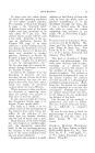
66 JOURNAL OF THE SOCIETY OF COSMETIC CHEMISTS eral nuclear remnants are also visible within the cortical cells. The second ultrathin section (Fig. 9) is interesting because it shows the way cuticle cells overlap. The ultrathin sectioning technique has one disadvantage in that the var- ious hair structures may not be adequately resolved. This is because the chemical composition of these structures is so similar that electrons may penetrate them to about the same extent, giving little contrast in the micrograph. Potentially, however, the ultrathin sectioning method shows much promise for studying hair by electron microscopic techniques. It has the advantage, as mentioned previously, of looking directly at the interior of the hair as it occurs in nature. DEGRADATION The third method useful for hair studies is the method of degradation to get separate particles of actual hair material. This method involves break- ing the hair down into its various histological components by a digestion, then examining the debris from the digestion in the electron microscope. Several reagents may be used for the digestion, for example, enzymes, acids and strong bases. Enzymes like trypsin are more suitable than strong acids or bases because they act more slowly and separate the hair into more distinct structures. This allows one to stop the degradation at any desired level of complexity and examine the resulting separate histological struc- tures. This method is useful for examining the separate components of normal hair and the effect of reagents upon these structures. It has a serious dis- -7- ¾ .'"• ..• ..: .. •4. 7 •--- •.. ':9 Figure 10.--Electron micrograph of fibril- lar material from a trypsin digestion of hair. Macrofibril is breaking down into micro- fibrils. Figure 11.--Electron micrograph of a scale (cuticle cell) fragment from a trypsin digestion of hair.

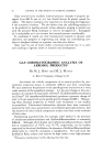
ELECTRON MICROSCOPE--TOOL FOR STUDY OF HAIR FIBERS 67 advantage in that one is never sure that any changes observed are due to treatment with reagent or due to the degradation process itself. A further difficulty is that the degradation products, in order to be shadow-cast for contrast, must be subjected to high vacuum. For that matter, the electron microscope itself is evacuated. Specific examples of the kinds of problems on which this method can be tried might be how the conformation of the cortical cells is effected by setting the hair with chemical agents or the pitting and etching of scale fragments caused by different reagents. As an example of the kind of pictures one can obtain, Fig. 10 shows a fragment of fibrillar material from a cortical cell at 10,000X magnification. In this micrograph, the fibrillar nature of the cortical cells is very evident. The larger fibrils at the right have been broken down far enough to see microfibrils. Figure 11 shows a fragment of a cuticle cell. Note the non- fibrillar nature of this as contrasted with the cortical cell. The proteinace- ous material within the cell has been partially removed from this one as evidenced by the many pits appearing in the surface. In the application of the various techniques which have been described to hair, it is always well to use both the light and electron microscope. The use of the ordinary light microscope can help one to better visualize what is being observed with the much higher magnification of the electron micro- scope. SUMMARY The three techniques of replication, ultrathin sectioning and degradation represent the most useful techniques for examining hair at the present time. I Their usefulness in any particular problem is limited only by the imagina- tion, skill and care of the individual applying the techniques. Often a com- bination of two or all three techniques must be used. It is certain that the techniques will be modified and improved and that new techniques for exam- iining hair in the electron microscope will continue to be developed as more I people become interested in the application of the electron •microscope to the solution of problems which may arise in the study of hair. REFERENCES (1) Barnes, R. B., Burton, C. J., and Scott, R. G., y. Atpplied Phys., 16, 730 (1945). (2) Manogue, B., and Moss, M. S., Nature, 172, 806 (1953). (3) Laxer, G., Sikorsi, J., and Whewell, H. J., Biochim. et Biophys. Atcta, 15, 174 (1954). (4) Birbeck, M. S.C., Mercer, E. H., and Barnicot, N. A., Exptl. CellResearch, 10, 505 (1956).
Purchased for the exclusive use of nofirst nolast (unknown)
From: SCC Media Library & Resource Center (library.scconline.org)