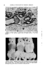
286 JOURNAL OF THE SOCIETY OF COSMETIC CHEMISTS effect on the pig's skin, except moderate hyperemia. The latter effect resembles more or less to the reaction produced on human skin. Clinical observations were made by Friederich (7), who treated 30 patients daily during three successive days over a 50 X 50 cm. area with 8 per cent ammonium thioglycolate solution. No changes could be ob- served in the blood pressure, sedimentation rate blood count, urine and liver function. Similar is the clinical observation of Behrman and Combes (8). It is of interest to note that 2 per cent thioglycolate has been used until recently to stabilize calcium gluconate solutions, without ever producing toxic effects. Although it is impossible to draw a final conclusion to humans from these observations, it is significant that Sulzberger and Rostenberg (9), as well as Ishikawa (10), were successful in sensitizing guinea pigs only with such substances which cause allergic reactions in humans. Thus Ishikawa was unsuccessful in producing hypersensitivity with nonsensitizing sub- stance, such as boric acid, and only with great difficulty' with bismuth which causes allergic reactions in rare instances. By analogy, one could postulate from our experiments that thrombocytopenic purpura, or periafter- iris nodosa is unlikely to be caused by either a commercial cold wave lotion, or a black hair dye. The failure to produce experimental thrombo- cytopenic purpura, or periarteritis nodosa, could also be explained by the well-known fact that sensitizing processes are relatively rarely proved in the etiology of both these diseases. It is just as illogical to link up leukemia with cosmetics, as it is without any firm foundation to consider the thioglycolates or paraphenylenedi- amine as a potential etiological factor of other blood dyscrasias. Millions use hair dyes and cold wave lotions without developing these diseases with measured frequency. Contrariwise, patients with this disease only rarely give the history of contact with the aforementioned cosmetics. In this connection, it is of particular interest to note that Bohrod (11) found this disease in an American deer and Stunzi (10) in 36 pigs. In the latter animals in five instances, erysipelas was considered a possible etological agent, and in 18 cases prophylactic vaccination against erysipelas. No other medicinal agent, either external or internal, has been used. Loc^L AND SvS'rEMtC ErrEc, or SEx HORMONES The second part of our presentation deals with the effect of percutaneous application of estrogens and androgens, on the sebaceous glands and hair growth in rabbits, as well as changes observed in the adrenal glands, most of which was published previously (13). It seems to me of interest to review the data together with some unpublished observations which might be of special interest to you. It should be briefly mentioned that 2 grams

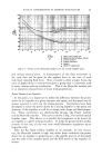
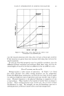
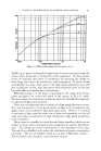
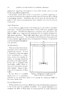
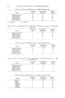
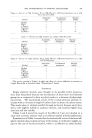
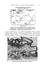
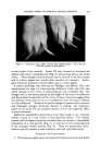
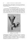
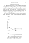
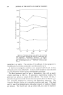
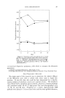
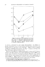
SYSTEMIC EFFECT OF TOPICALLY APPLIED COSMETICS 287 of ointment were applied between eighteen to twenty-four weeks. Con- trol rabbits were similarly rubbed with 2 grams of the ointment base. Each gram of ointment contained 0.15 mg. of alpha-estradiol, and 2 rag. methyltesterone. At intervals of three weeks the abdominal skin of the rabbits were shampooed and the border surrounding a twenty-four square inch area was clipped and the hair discarded. After this, the hair from the twenty-four square inch area was closely clipped and dried to a con- stant weight in an oven at 56øC. Twenty-one rabbits, partly castrated, partly noncastrated, were used in this study. Several clippings, varying in number from three to twenty, were made and the weights averaged. Discussion OF THE EFFECT OF ESTROGENS AND ANDROGENS It was clearly demonstrable in this experiment that estrogenic hormones produced an increased activity of the hair papillae and reduction of the size of sebaceous glands and also a stimulation of hair growth. This was evident in increased weight of clipped hair in contrast to the lesser weight of an equal sized clipped hair in the control animals and in those animals which were treated with male sex hormones (Fig. 1). The fact that the increase of hair occurred not only on the inuncted area, but on the opposite side as well, indicates a general, rather than a local effect (Fig. 2). In this experiment castrated and noncastrated rabbits were used, and we not only could demonstrate the same phenomenon in both group of animals, but also the reversibility of the estrogenic effect by subsequent applica- tion of male sex hormones (Fig. 4). It is of interest to note that castrated male rabbits treated with the ointment base showed a similar histological pattern, as the male rabbits revealed after applications of progynon oint- ment (alpha estradiol) (Fig. 3). It is apparent that the absence of male gonads caused an appreciable reduction of the size of sebaceous glands which is generally associated with an increased growth of hair. The latter findings correspond with the observations made by Hammilton (14, 15), Roony and Zakon (16, 17) on humans. According to Hammilton, andro- genic hormones are usually implicated in extensive baldness, but alopecia does not develop in eunuchs and in those men who fail to mature sexually. Roony and Zakon also demonstrated microscopically that methyltestos- terone stimulates the sebaceous glands of males and that diethylstilbestrol has a depressive effect the sebaceous glands of the adult male. Our findings were confirmed by Light (18) in a brilliantly executed study on rats. He stressed especially the anabolic effect of androgenic hormones to cause connective tissue infiltration of the vascular bed about the hair roots which are supposed to interfere permanently with the immediate nutrition of hair follicles. Our histological studies on rabbits did not reveal similar changes. Although we did not include biochemical data, which would corroborate
Purchased for the exclusive use of nofirst nolast (unknown)
From: SCC Media Library & Resource Center (library.scconline.org)