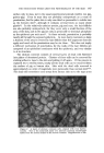
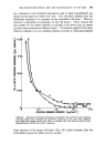
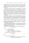
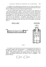
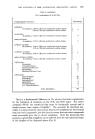
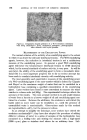
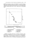
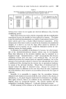
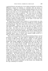
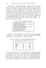
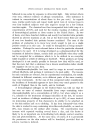
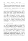
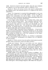
THE STRUCTURES WHICH LIMIT THE PENETRABILITY' OF THE SKIN 147 surface only in man, not in the usual experimental animals (rabbit, rat, pig, guinea-pig). Even in man they are probably unimportant as a route of penetration, for the palm skin is only one-third as permeable to iodide ions as the forearm skin n, although it contains several times as many sweat glands •ø'. In the relatively unhairy species, pig and man, the hair follicles are also probably unimportant, for they cover only a small fraction of the area of the skin, and in the pig are only as permeable to trinbutyl phosphate as the epidermis per unit area •8. In these animals, penetration is probably nearly all through the exposed epidermis. In more hairy mammals so large a fraction of the area is covered by hair follicles that this argument does not apply and much of the penetration may be via them. This does not imply a different mechanism of penetration, for the walls of the hair follicles are composed of an epithelium continuous with the epidermis, and very similar to it in structure. The stratum corneum consists of several layers of dead cells flattened into plates of denatured protein. Clusters of these cells can be removed by sticking adhesive tape to the skin and pulling it off again. If this process is repeated ten to twenty times, nearly all the dead cells can be removed from the surface of pig or human skin. Skin with the dead cells removed is approximately an order of magnitude more permeable than normal skin •4'•*. The dead cells sometimes come away from human skin on to the tape as an ß ' ':"• --: :':• "• 'ii•.-' ,,• ? i. •?:::'•!:.•:'-' ?': '-'-:i .•'•i :' '" " .... F' ::"• g•:i : •' .• •...[•:: • •:• ...•-: ' . • :.• ..- . •.• ::• . ...5 ' '.• .-,::• .... . .• :. ..:•: .•. •-' ß • "•. .•.• .•" .. :? " • '.• :.. :.•...::... " .. .• .: ::.• .:.. .[.. •... • .: .•: • .:.-......• ß •': •.•...•:•...•.. '.:' •... -... ...... .. .:• ..x: ..• • . ....:..•, .• , , .., .: • ..... Figure •. A layer of stratum corneum cells, stained •th iron haemo- toxylin. This is the innermost layer of a stratum corneum membrane •ø •ken from the doral surface of the human fore•m. Magnffication x 500.

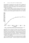
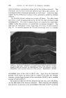
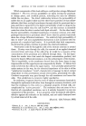
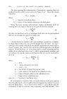
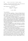
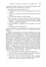
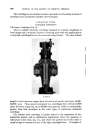
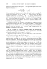
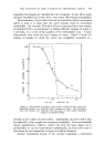
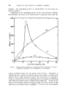
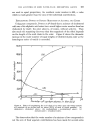
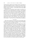
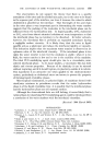
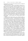
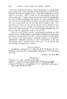
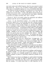
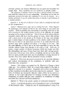
148 JOURNAL OF THE SOCIETY OF COSMETIC CHEMISTS intact membrane, representing a large part of the stratum corneum •6. The membrane can be removed from the adhesive tape with a latex solvent. It is as impermeable to trinpropyl phosphate as whole skin •7. Both these facts suggest that the diffusional resistance of the epidermis is within the stratum corneum. The stratum corneum contains ten or more cell layers. The cells of each layer form a crude hexagonal lattice (Fig. oe) with the edges of adjacent cells interdigitated xS. The cells of adjacent layers are connected by small inter- cellular bodies (desmosomes) of considerable mechanical strength •9. The cells have an osmiophilic border, suggesting some form of cell membrane (Fig. $). In sections prepared for electron microscopy there is a definite .Figure 8. Electron micrograph of a vertical section through the stratum corneum of human thigh skin. Note the osmiophilic borders of the cells, the variable density of the cell contents, the interdigitation of the cell processes, and the intercellular bodies (poorly preserved in this section). Magnification x 20,000. intercellular space of the order of 100 A wide. Apart from the denatured protein, which makes up about half the weight of the cells, the stratum corneum contains large amounts of free aminoacids (10-20%) and a variable amount of lipid (2-20%) 3ø. The clusters of cells torn off with adhesive tape are only a few cells thick, not the full depth of the stratum corneum •x. Each stripping with adhesive tape produces an increase in the permeability of the skin to ions • (as shown
Purchased for the exclusive use of nofirst nolast (unknown)
From: SCC Media Library & Resource Center (library.scconline.org)