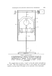
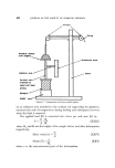
THIN-LAYER CHROMATOGRAPHIC TECHNIQUES IN RESIDUE ANALYSIS 475 the observed R F value of the chromatographed pesticide usually a reduc- tion of this value is noticed. Organo-chlorine pesticide residues after extraction from animal produce have been cleaned-up for gas-liquid chromatographic examination by means of a dimethylformamide (DMF) partition process, followed by passage through an alumina or magnesia column (42). A similar clean-up process is suitable for thin-layer chromato- graphic purposes but the columnar clean-up stage may usually be omitted. Similar partition processes using acetonitrile (43) and dimethylsulphoxide (44) have also been used for this purpose. In general it is the total load of material that is to be applied to the chromatoplate at one point that governs the thickness of the layer required for efficient chromatographic clean-up. For clean extracts the usual layer thickness of about 250[• is adequate for preparative and clean-up purposes 500• or 1 mm thickness is more usual as previously described. The amount of previous clean-up required may therefore be related, in part, to the availability of spreading apparatus capable of producing layers of various thickness. Application of the sample extract In order to obtain accurate and reproducible thin-layer chromatograms a few simple rules must be observed during the application of the sample extract to the adsorbent layer. The nature of the solvent used for the final solution of the extract to be applied to the plate is very important. In order to ensure that the size of the spots shall be as compact as possible, the chosen solvent should be of as low a polarity as is consistent with good solubility of the pesticide. This is particularly important when only very dilute solutions are available, when it may be necessary to apply the extract repeatedly to one spot in order to ensure sufficient material for dear visualization. Such "over-spotting" is liable to induce radial chromato- graphy at the origin when polar solvents are involved. The solvent should also be readily volatile yet not of so low a boiling point that standard solutions cannot be maintained in constant concentration or that evapora- tion, with consequent deposition of the dissolved material, occurs while within the applicator. Solvents with boiling points in the range 40-60øC are usually preferred whenever possible. The sample solution may be applied to the layer surface by gently touching it with a filled calibrated capillary or micro-pipette or by means of a micro-syringe of suitable capacity volumes of the order of 1 to 15 [•1 per spot are preferred. To aid in the accurate alignment of a series of

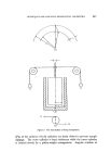
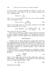
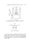
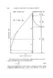
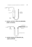
476 JOURNAL OF THE SOCIETY OF COSMETIC CHEMISTS spots across the chromatoplate, a suitably drilled template, usually con- structed of a transparent plastic material, is often used both as a guide and to protect the surface of the layer. The line of spots so applied should be about 15 to 20 mm from one edge of the carrier plate and parallel to it, individual spots being at least 1 cm apart. The line should also be per- pendicular to the direction of spreading to avoid possible unevenness in thickness across the layer. Care must be taken not to penetrate the layer with the tip of the applicator or irregular shaped spots are liable to result on developing tlxe chromatogram (45). In order to maintain the desired compactness (0.5 cm) of the origin spots, Mangold (27) advised the use of a stream of nitrogen to ensure rapid evaporation of the solvent from the plate, while Miller and Kirchner (46) employed a low temperature hot-plate for the same purpose. Ritter and Meyer (47) have mechanized the operation of spotting the samples on to the layer. They designed a syringe which traverses the chromatoplate, applying controlled doses of the solution as required a similar "streak" applicator was used by Coleman (48). Morgan (49) pro- duced a multiple capillary device for the simultaneous application of many solutions to a single chromatoplate Bark et al (50) found that such rapid spotting techniques improved R F reproducibility. As alternative modes of spotting, Metz (51) has used a system of elution from a filter paper triangle, while Tate and Bishop (52) described the use of a wire loop such as is employed for bacteriological purposes. Although the application of circular spots is most often recommended, Honegger (53) advocated that the sample should be applied as a thin band, about 1 cm long. Techniques of this nature are especially suitable for extracts of residues which have not undergone a very thorough clean-up process and in which the ratio of co-extractives to residue is rather high. The load of material which may be successfully applied to the chromato- plate is limited by the layer thickness on one hand and by the sensitivity of the visualization system on the other. Generally speaking, amounts of material in the range 0.5 to 10 [•g may be chromatographed on 250• thick layers to give clearly defined regular spots with little or no streaking. Where large amounts are encountered it is advisable to dilute the extract further and to repeat the chromatogram. Variable R F values have been observed where overloading of the adsorptive capacity of the layer has occurred (54). In the case of purely preparative thin-layer chromato- graphy, however, this factor is frequently less important than in diagnostic work and large loads may then be safely used.
Purchased for the exclusive use of nofirst nolast (unknown)
From: SCC Media Library & Resource Center (library.scconline.org)