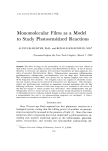
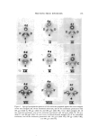
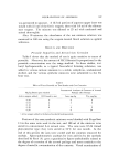
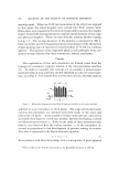
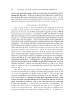
172 JOURNAL OF THE SOCIETY OF COSMETIC CHEMISTS for 15 minutes. The pH of the supernatant was lowered with HC1 to 4.5 at which pH maximum fiocculation of the alkali-tonofibrin deriva- tives occurred. The protein precipitate was centrifuged at 1500 rpm for 5 minutes, redissolved by the addition of dilute alkali, and reprecipitated at pH 4.5 in the same manner as above. After this procedure of solubili- zation and reprecipitation was repealed again, the proteins were dissolved in barbital buffer, pH 8.6. The protein content of the various solutions of epidermal proteins was determined directly by drying the samples at 110øC overnight and then subtracting the weight of the barbital buffer. Double diffusion analyses were carried out on microscope slides in 1.5% agar buffered with barbital, pH 7.5 (/• = 0.15). The agar also con- tained 7.5% glycine which was found to help maintain the solubility of the antigens. Sodium azide (0.1%) was added as a preservative. Specific absorptions of all the antisera with human serum and then with the various antigens were accomplished by first delivering the antigens and serum into the well of the agar gel. This was followed by addition of the various antisera. This process is hereafter referred to as pretreatment of the antisera as is indicated in the figure legends. After application of the antigen solutions and antisera, the slides were developed in a moist at- xnosphere for one week. These slides were then washed with saline, one hour with distilled water, covered with filter paper strips, and dried. Buffalo black was used to stain the precipitin bands. RESULTS In Figs. 1-4 the following letters and numbers were used: D, E, and F for the urea, alkali, and tonofibrin-derived antigens, respectively 4, 5, and 6 for the antisera prepared against D, E, and F, respectively and d, e, and f for the antigens (same as D, E, and F) except as used for pretreat- ment of the antisera. For example, 4d would indicate antiserutn pre- pared against the urea antigens and pretreated with the same antigens 4f would denote antiserum prepared against the urea antigens and pre- treated with the tonofibrin-derived antigens, and so on. In Figs. 1-3, the various antigens D, E, and F were placed in the side wells, the untreated antisera 4, 5, and 6 in the lower wells or below the antigens, and the pre- treated antisera 4-d, e, f 5-d, e, f and 6-d, e, f in the upper wells or above the antigens. Pretreatments are indicated in vg protein for 20 •1 of the various treated and untreated antisera. The amount of each antigen used in the side wells was for D, 184 vg E, 88 vg and F, 180 vg protein. Anti-

PROTEINS FROhi EPIDERMIS 173 4e Figure 1. Agar gel precipitation patterns of the antiserum prepared against the urea antigens verstts urea antigens (D), of the untrcatcd antiserum, and of the antiserum pretreated with 184 txg d (slide I), 440 txg e (slide II), and 360 txg f (slide III) versus alkali antigens (E), of the untreated antiserum, and of thc antiserum pretreated with 184 txg d (slide IV), 88 txg e (slide V), and 360 txg f (slide VI) and versus to•:ofibrin-derived antigens (F), of the untreated antiserum, and of the antiserum pretreated with 184 t•g d (slide VII), 176 txg e (slide VIII), and 180/•g f (slide IX)
Purchased for the exclusive use of nofirst nolast (unknown)
From: SCC Media Library & Resource Center (library.scconline.org)