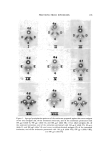
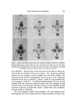
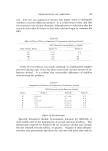
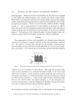
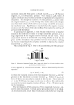
180 .JOURNAL OF THE SOCIETY OF COSMETIC CHEMISTS quite strongly with the urea, alkali, and tonofibrin-derived antigens. From the results given in Fig. 4, slides I-III, it is apparent that the an- tisera prepared against the urea and alkali proteins gave a more complex reaction with the various proteins than did the antiserum prepared against the tonofibrin-derived proteins. The antisera against the urea and alkali proteins yielded, with the three antigens, one and three pre- cipitin bands of identity, respectively, and the three antigens reacted strongly with the antisera prepared against the three antigens. It is ap- parent then that although the urea and alkali proteins have some different physical properties (3), they are either related immunochemically or that 6M urea and dilute alkali extracted similar proteins, but in different aInounts. For example, the antisera prepared against the alkali, urea, and tonofibrin-derived proteins showed, respectively, 3, 3, and 2 bands of pre- cipitation with the urea antigens 4, 2, and 2 bands of precipitation with the alkali antigens 3, 3, and 2 bands of precipitation with the tonofibrin- derived antigens. In other words, the reactions of the three proteins were quite strong with each of the antisera prepared against them. Antiserum prepared against the proteins extracted from epidermis with dilute alkali had precipitation bands of identity with the other two protein prepara- tions (Fig. 4, slide II), even though the reactions were quite weak with the nrea proteins. In this instance, the urea antigens had a strong indepen- dent precipitin band such was also the case for the reaction of the urea antigens with the antiserum prepared against the urea antigens (Fig. 4, slide I). Hence, it appears that urea, dilute alkali, and lithium bromide extracted several similar proteins in different amounts or, less likely, that these proteins are related. The alkali and tonofibrin-derived proteins ap- peared to have more in common than the urea proteins (Fig. 4, slides An interesting difference between the urea and alkali proteins is the following observation. Antiserum prepared against the urea proteins pretreated with alkali antigens failed to inhibit precipitin band formation with the urea antigens (Fig. 1, slide II), and antiserum prepared against the alkali antigens pretreated with urea antigens failed to impede precipi- tin band formation with the alkali and tonofibrin-derived antigens (Fig. 2, slides I and IV). In line with these differences, antisera prepared against the alkali and tonofibrin-derived proteins reacted weakly with the urea antigens. These observations on the various antisera and antigens re- sulted froxn the fact that alkali extracted more of some proteins than did m-ea and that urea extracted several proteins in a relatively uniform man-

PROTEINS FROM EPIDERMIS 151 her. Although tono[ibrin-derived proteins have a different source (tono- fibrin) and an isoelectric point (pH 4.5) different from that of the urea and alkali proteins (pH 5.5), their immunodiffusion patterns were quite similar. These complex interrelationships are under further study. Ac- cording to electrophoretic and schlieren patterns (sedimentation) of the urea and alkali proteins of mouse epidermis, normal and pathological (3), and of human epidermis (13), these proteins appeared homogenous. The alkali and urea proteins of mouse epidermis differ significantly in such properties as viscosity increment, axial ratio, frictional coefficient, sedimentation constant, and molecular weight (3) the sedimentation constants of the urea and alkali proteins of human epidermis are also dif- ferent (3). However, these differences in physical properties may be due to varying' sizes of the same proteins extracted by the different reagents. Since the urea proteins (epidermin) of Rudall (1) and tonofibrin of Roe (7, 8) may be involved in keratinization (according to these investigators), further work is necessary to determine the relationships of the alkali and tonofibrin-derived proteins to keratinization. ACKNOWLEDGMENT The skilled assistance of Miss A. Baumler is gratefully acknowledged. (Received August 8, 1969) REFERENCES (1) Rudall, K. M., The proteins of mammalian epidermis, Advan. Proteitz Chem., 7, 253- 90 (1952). (2) Carruthers, C., •,Voernley, D. L., Baumler, A., and Kress, B., Proteins of mammalian epidermis, J. Inve, t. Dermatol., 25, 89-101 (1955). (g) Carrnthers, C., Woernley, D. L., Baumler, A., and Lilga, K., Studies on certain proteins in normal and pathological epidermis, Brit. J. Cattcer, 11, 597-604 (1957). (4) Carruthers, C., Woernley, D. L., Baumler, A., and Shorts, H., The strnctural proteins of epidermis and their possible relation to aging skin, J. Soc. Cosmetic Chemists, 6, 324-43 0955). (5) Crounse, R. G., Epidermal kerrtin: A re-evaluation, Nature, 201), 539-42 (1963). (6) Mercer, E. H., and Olofsson, B., Sedimentation analysis of an extract of tfie prekerati- nous layers of skin, J. Polym, er Sci., 6, 261-9 (1951) . (7) Roe, D. A., A fibrous keratin precursor from the human epidermis. I. The extrac- tion and physical properties of a fibrous protein fmtnd in the human epidermis, I. In- vest. Dermatol., 27, 1-8 (1956). (8) Roe, D. A., Further studies of a fibrous keratin precursor from the human epidermis, Ibid., 27, 319-24 (1956). (9) Rothberg, S., The synthesis of epidermis proteins. I. Alkali solnbilized (insolnble) protein, Ibid., 4•, 151-7 (1964). (10) Rothberg, S., The synthesis of epidermal proteins. II. Solnble proteins, Ibid., 4,% 159- 64 (1964).
Purchased for the exclusive use of nofirst nolast (unknown)
From: SCC Media Library & Resource Center (library.scconline.org)