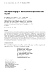
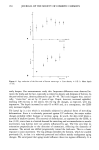
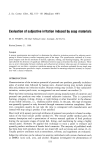
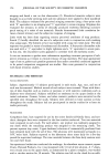
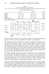
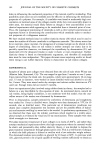
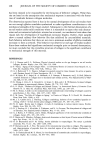
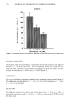
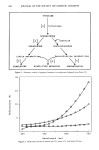
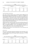
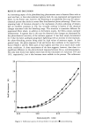
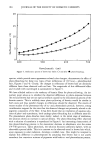
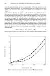
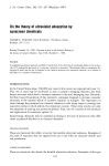
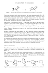
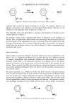
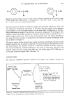
150 JOURNAL OF THE SOCIETY OF COSMETIC CHEMISTS age (years) E •- •o =o 2'0 .'o 6'ø e•o ' age (years) Figure 4. Leg: evolution ooe the first axis ooe oeurrow versus age. a. Line density ___ S.oe. b. Mean d½lxh --- S.E. sarily deepen. Our measurements verify this. Important differences were observed be- tween the limbs and the face, especially as related to density and deepness of furrows. In the peri-orbital area, density plateaued at age 30-40. This could suggest that, statisti- cally, "crows feet" are set by 35 years of age. Depth, however, increased eightfold, reaching 200 microns in the sixties. On the leg the changes, as expected, were less impressive. The depth increased by only 65 to 80% and, as a consequence, the CDSS only increased slightly. The lateral leg is a site which is minimally subjected to physical forces of stretching deformations. Since it is relatively protected against UV radiation, the major surface changes probably reflect biologic or intrinsic aging. In youth, the skin relief shows a network of shallow furrows. The reservoir of deformation, as expressed by the CDSS, is low (1.02), since there is a limited demand for stretching and accommodation to muscle movements. Leg furrows were not greatly influenced by age. The first axis kept its orientation perpendicular to the direction of constraints, reflecting the dynamics of extension. The second one shifted progressively toward the limb axis. This is a classic response to joint movement. The leg perhaps resembles the forearm, which we studied previously (5), in that it is relatively protected and reflects mainly endogenous aging changes. We anticipated that aging would influence these two regions differently. It is

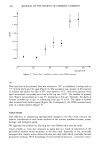
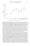
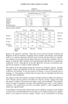
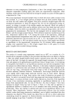
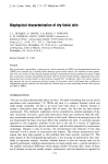
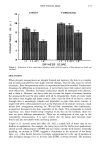
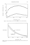
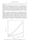
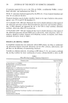
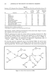
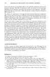
AGING AND MICRORELIEF OF SKIN 151 I .1 0 I .05. 1.00. 0 20 40 60 80 Figure 5. Leg: evolution of the CDSS versus age. age (years) informative to compare these changes to the volar forearm which is submitted to large amplitude deformations such as flexing, extension, and rotation. Like the leg, it is a relatively protected area. The different evolution of the skin relief in these two sites can be attributed to their being subjected to different mechanical stresses. The main aging change in the forearm's topography was a shifting of the primary lines towards the limb axis, increasing anisotropy. This also occurred in the leg, but to a lesser degree. On the forearm, accordingly, the reservoir of deformation, needed to accomodate these physical movements, was relatively high in the young (CDSS - 1.16). Repeated stresses throughout life necessarily bring about some reorientation of the furrows, no- tably a shift of the major lines towards the long axis of the limb, accompanied by a progressive loss of the second axis. Despite the latter and the decreased density of furrows, the CDSS actually increased owing to deepening of the furrows. Finally, it should be emphasized that the changes in orientation and depth of the major furrows with aging reflect alterations in the underlying dermal matrix. In this respect, deterioration of the elastic fiber network has greater impact than structural disorganiza- tion of collagen. Elastic fiber degeneration is noticeable at the ultrastructural level as early as age 30 when cavitations appear even in unexposed buttock skin (8). Likewise in protected skin, the subepidermal vertical skeins of fine elastic fibers, mainly bundles of microfibrils, gradually disappear and are completely lost by age 50 to 60. These events lead to laxity and loss of elastic rebound so that the skin becomes more vulnerable to mechanical stresses. When, as on the face, prolonged exposure to sunlight virtually destroys the elastic network, the skin becomes even looser and more prone to folding, being greatly in excess. Loss of collagen in elastotic tissue adds to this superabundant, loose, recoiless, flabby skin. In protected skin, changes in collagen are far less conspic- uous and of lesser importance compared to elastin. With aging, collagen becomes stiffer
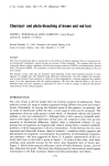
Purchased for the exclusive use of nofirst nolast (unknown)
From: SCC Media Library & Resource Center (library.scconline.org)