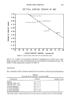
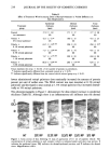
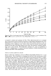
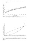
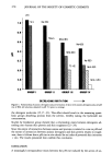
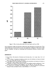
j. Soc. Cosmet. Chem., 39, 235-240 (July/August 1988) The effect of retinyl palmitate on skin composition and rnorphornetry DAVID F. COUNTS, FRANK SKREKO, JULIANNE McBEE, and A. G. WICH, The Lilly Research Laboratories, Eli Lilly and Company, Lilly Corporate Center, Indianapolis, IN 46285. Received January 21, 1988. Synopsis Topical administration of increasing doses (0.1%-5% (w/w)) of retinyl palmirate (RP) for 14 days, in a suitable cosmetic vehicle, caused significant dose-related changes in skin composition and morphometry of the hairless mouse. There was a maximum 32% increase of protein per unit of skin surface area and a maximum of 128% increase of collagen per unit of skin surface area in response to RP administration when compared to control vehicle. In addition, there was an increase of DNA content in response to RP adminis- tration. There was significant thickening of the epidermis in response to the increasing dose of RP. Al- though the total thickness of skin was not significantly increased by RP application, the total skin thick- ness was greater than the untreated control or the control-vehicle-treated animals at every dose of RP tested. These results indicate that the topical application of RP (in an active form) can alter the composition and morphometry of the skin in the hairless mouse. INTRODUCTION Vitamin A or retinol is essential for normal skin development. Vitamin A is an impor- tant regulator of keratinocyte terminal differentiation (1). An excess of vitamin A in- hibits keratinization (1), whereas a deficiency results in squamous metaplasia and kera- tinization of epithelial tissue (2). Thus, epidermal development is, in part, regulated by vitamin A. It is also known that vitamin A can alter or modulate total collagen syn- thesis (3-5). Also, retinoic acid has been demonstrated to alter the type of collagen synthesized (6). Retino! has the potential to alter the expression of protein molecules in both the epi- dermis and dermis. Although retinoic acid can alter both keratinocyte as well as fibro- blast (7) protein metabolism, the precise effect of retino! on intact skin composition has not been determined. Several cosmetic formulations contain vitamin A. The present study was undertaken to determine whether or not vitamin A, if present in an active form in cosmetic formulations, can alter skin composition and morphology. MATERIALS AND METHODS ANIMALS Female hairless mice (HRS/J) (six to eight weeks old) were obtained from Jackson Labo- 235

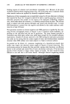
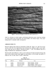
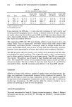
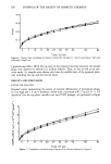
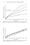
236 JOURNAL OF THE SOCIETY OF COSMETIC CHEMISTS ratories (Bar Harbor, Maine) and maintained for one week prior to all studies. The mice were housed five animals per cage in standard "shoe box"-type cages (6 in x 10 in x 5 in) and given food and water ad libitum. Each animal received 0.1 ml of a test cosmetic formulation applied to the dorsal skin surface and rubbed into the skin (approximately 5-10 sec), using a gloved finger as an applicator. This treatment was continued each day for 13 additional days. BIOCHEMICAL AND HISTOLOGICAL ANALYSES On Day 15, the animals were terminated by cervical dislocation and the skin removed from the dorsal, treated surface of the animals. One section of the skin was taken for histological evaluation and a second piece was scraped free of subcutaneous fat. Two circular punch biopsies (6 mm in diameter) were taken from the latter, blotted dry, and weighed. These biopsies were then pulverized in liquid nitrogen and homogenized in phosphate-buffered saline using the Polytron-ST homogenization system. This homoge- nate was used to determine the protein content, DNA content, and collagen content (as measured by proteinaceous hydroxyproline content of the tissue) (8). An equal volume of 10% (w/v) trichloroacetic acid (TCA) was added to the homogenate, and the suspen- sion mixed and allowed to equilibrate for five minutes (3-5øC). The suspension was centrifuged at 10,000 x g for ten minutes. The pellet was suspended in 0.5 ml of 5% (w/v) TCA and centrifuged at 10,000 x g for ten minutes. The pellet was suspended in 0.2 ml of 10% (w/v) TCA and heated at 90øC for 20 minutes. The suspension was then set in an ice bath for 30 minutes. The suspension was then centrifuged at 10,000 x g for three minutes. The supernatant was analyzed for DNA as described by Schneider (9). The precipitate was suspended in 0.5 ml of 0.5M NaOH, heated for one hour at 55øC, and an aliquot was removed for protein determination (10). The remainder of the sample was sealed in a glass ampule in vacuuo with an equal volume of 12 N HC1 and hydrolyzed for 24 hours at ! !0øC. After hydrolysis the sample was dried in a desiccator and the dried material was assayed for hydroxyproline (! 1). The skin samples taken for micromorphometric analyses were fixed in buffered neutral formalin solution, dehydrated, and embedded in paraffin blocks which were then cut into 7-1•m-thick sections. These sections were stained with hematoxylin and eosin for later examination. Thickness measurements were based on the cross-sectional thickness of the skin. Several (six to eight) sections were stained and used for evaluation per animal. Thickness measurements (eight to ten) per animal were made by selecting the measurement site at random. The average of these measurements was considered as the measurement for one animal. Total skin thickness was considered the thickness of the skin from the outer stratum corneum to the panniculus carnosus. Dermal thickness was taken as the distance from the dermal epidermal junction to the top of the dermal adipose deposits. COSMETIC FORMULATION All cosmetic formulations used in this study were oil-in-water emulsions and were manufactured under a nitrogen blanket. The water phase of the emulsion systems ranged from 79-$4%, the oil phase was !6%, and the retinyl palmitate (Roche) con- centration ranged from 0-5 %. Water was removed from the emulsion to accommodate
Purchased for the exclusive use of nofirst nolast (unknown)
From: SCC Media Library & Resource Center (library.scconline.org)