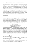
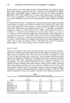
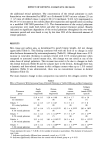
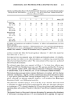
ANDROGENS AND PROPIOBACTERIAL ENZYME ON SKIN 261 Table I Individual and Mean Daily Doses ---SE of Self-Administered Testosterone and Anabolic Steroids (mg/day) During the Four-Week Periods (I = 0-4 wk, II = 5-8 wk) in Seven Power Athletes Between the Collections of Bacterial Samples Subjects 1 2 3 4 5 6 7 mean -+ SE Testosterone I (0-4 wk) II (5-8 wk) Nandrolone I (0-4 wk) II (0-8 wk) Methandienone I (0-4 wk) II (5-8 wk) Stanozolol I (0-4 wk) II (5-8 wk) 23 7 19 19 26 23 15 19 -+ 2.4 27 9 23 23 17 24 25 21 --- 2.3 19 7 7 7 5 5 6 8 --- 1.8 26 14 7 7 3 5 8 10 --- 3.0 21 - 13 13 15 10 10 14 _____ 1.5 30 - 20 20 20 10 20 20 - 2.4 19 2 2 - - - 8 _+ 3.9 13 2 2 - - 7 6 + 1.8 All athletes used testosterone and nandrolone. In addition, six athletes used methandienone, and three athletes stanozolol. Trivial and systemic names: testosterone, 1713-hydroxyandrost-4-en-3-one nandrolone phenylpropionate, 1713-hydroxyestr-4-en-3-one 3-phenylpropionate methandienone, 17or-methyl 1713-hydroxy- 1, 4-andro- standien-3-one stanozolol, 17-methyl-2H-5ot-androst-2-eno[3,2-C]pyrazol-1713-ol. activity is noted only when the bacteria produce reductase (+) above the sensitivity level of this test or not (-). The lipase activity was measured using the Unkles and Gemmel method (15). Briefly, lipase activity was visible on the tributyrin plates as a turbidity ring around the hole where bacterial lipase had been added. The measurements of the rings were performed after 24 hours of incubation. The tributyrin agar plates were prepared as follows. 4.7 g brain heart infusion agar (Oxoid) and 100 ml of distilled water were used. The agar was sterilized for 15 mins at 12 IøC and then cooled to 56øC, after which an emulsion of tributyrin containing 4 ml of distilled water, 5 Izl of Tween 80, and 1.0 ml of tributyrin (Fluka) was added. The hyaluronidase assay agar plates contained hyaluronic acid sodium salt from human umbilical cord (Sigma), 2 mg/ml, and the condroitinase assay plates chondroitin sulfate C, super special grade from shark cartilage (Seikagaku Kogyo Co., Ltd), 2 mg/ml. Both plates contained 5% albumin (Albumin Fraktion V aus Rinderserum (Boehringer Mannheim Gmbh) ) and 2000 Izg/ml penicillin. The plates were used after drying at 37øC on the day of use. Fourteen holes were bored in each plate into which the bacterial samples pipetted. After this, the plates underwent an incubation at 37øC in humid chambers for 24 hours for tributyrin and hyaluronidate specimen plates and another 24 hours for tributyrin Tween 80 and chondroitin sulfatase plates. The bacterial density of the growth tubes was determined by measuring the absorbance of well mixed culturing solution at 45 nm immediately after applying the duplicate

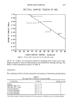
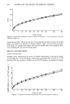
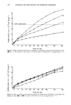
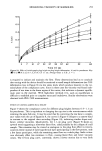
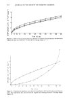
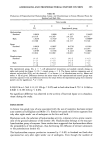
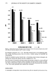
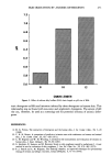
262 JOURNAL OF THE SOCIETY OF COSMETIC CHEMISTS enzymic specimens. The growth density was read from a standard curve, which was made by serially diluting a dense bacterial suspension with BHI solution and then measuring the absorbance of each dilution at 450 nm. The densest bacterial suspension is represented as 100% and its dilution as 75%, 50%, 25%, etc. From the curve (absorbance as a function of the density percentage) it was possible to read the per- centage density of each culturing tube. Dividing the turbidity value by the percentage mentioned above, a comparison unit per hole (U/h) was achieved (clearing zone diameter in mm per bacterial growth density), which describes the activity of enzyme production. These procedures were performed during weeks 0, 4, and 8 for the experimental group and once during week 0 for the control group. For ethical and legal reasons these effects could not be investigated according to the strict rules of controlled clinical trials but only within a given sports environment. Means and + SE of means were calculated. Differences between mean values of the experimental and control groups were tested by unpaired t-test, and differences between the values within the experimental group were tested for significance by paired t-test. RESULTS Table II shows that the control subjects were smaller than the athletes in the experi- mental group but had more body fat in relative terms. In the experimental group, there was weight gain during androgen administration but no increase in body fat. Testicular volume decreased during androgen administration by 35.9% (16), from 21.4 + 1.4 ml to !3.7 - 0.9, respectively. Four out of seven experimental subjects developed 35-50 papulopustular lesions on the upper trunk and face but only after eight weeks' use of testosterone and anabolic ste- roids. At the beginning of the study there were no differences in the production of P. aches enzymes between the experimental and control groups (Table III). Only after eight weeks' use of androgen steroids did hyaluronidase production increase in forehead skin and back skin strains. The increase in forehead skin was from 1.000 + Table II Anthropometric Characteristics of the Group Studied Body weight (kg) Body fat (%) Testicular volume (ml) 90.9 + 9.2 11.1 -+ 1.0 21.4 + 1.4 75.3 -+ 4.7*** 13.5 -+ 0.0 NS - EG 96.5 + 10.5'** 10.8 + 2.6 NS 13.7 + 0.9*** At the start of the investigation At the end of the investigation EG CG EG = experimental group, n = 7, self-administered testosterone and anabolic steroids during the eight- week study. CG = control group n -- 8. The values indicate means + SE, NS = not significant, *p 0.05, ***p 0.001 (two-tailed Student's t-test).
Purchased for the exclusive use of nofirst nolast (unknown)
From: SCC Media Library & Resource Center (library.scconline.org)