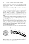
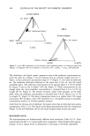
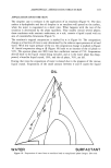
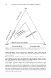
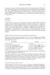
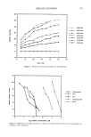
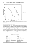
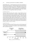
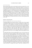
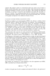
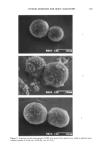
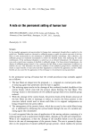
190 JOURNAL OF THE SOCIETY OF COSMETIC CHEMISTS dorsal back skin using a 1-cc syringe and spread evenly with a glass rod (sunscreen) or a Pipetman © (Rainin Instrument Co., Woburn, MA) and spread evenly with the flat side of the pipet tip (ot-tocopherol and iron chelators). When groups were treated and irra- diated, treatments were applied two hours prior to UVR exposure except for sunscreen treatments, which were given immediately prior to exposure. For combination che- lator-plus-sunscreen treatments, the chelator was applied two hours before UVR expo- sure, followed by the sunscreen applied immediately before exposure. After treatment, mice were returned to their cages without restraint until irradiation. EXPERIMENTAL PROTOCOL The protocol used in this study is shown in Figure 1. Groups of hairless mice were subjected to a UVR exposure regimen (3 X/week) for 15 weeks without topical treat- ment. The UVR sources were either a solar simulator or UVB fluorescent tubes. After the UVR pre-exposure period, topical treatments were begun (3 x/week) with the pho- toprotective agents. During the treatment period, some groups of mice were continued on the UVR exposure regimen (continued UVR), while others were treated without further irradiation (stop UVR). After the treatment period (12 or 20 weeks), total tumor area and basal epidermal ODC activity were measured. This protocol mimics the probable human situation of some level of solar damage to the skin before the first use of photoprotective product. The purpose of either continuing or discontinuing irradiation after starting treatment is to mimic the human situation of either continued further sun exposure or avoidance of further sun exposure. TUMOR QUANTIFICATION Skin lesions were diagnosed as tumors if they were round, red, raised, and greater than 1 mm in diameter. Total tumor area per animal was accurately quantified from photo- graphic transparencies of the backs of the animals by using a computerized digitizing tablet as previously described (28). Continue UVR Exposure I Stop UVR , Exposure ' No Treatment Photoprotective Period Treatment Period I I 5 10 15 20 25 30 35 Time (weeks) Figure 1. UV irradiation and treatment protocol.

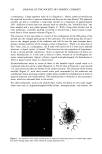
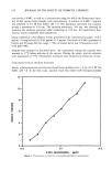
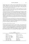
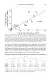
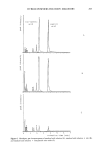
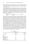
UV-INDUCED SKIN TUMORS 191 ASSAY OF ODC ACTIVITY Mouse epidermal ODC activity was determined as previously described (18). For the assay of human epidermal ODC activity, epidermal shave biopsies (• 1 cm 2) were ob- tained using a double-edged razor blade, the outer edges covered with cellophane tape, leaving the desired 1-cm cutting edge exposed [34]. Anesthesia for biopsy was achieved by applying an ice cube to the skin for two minutes prior to biopsy. Biopsies were immediately transferred to a tube containing 0.6 ml ice-cold homogenization buffer [50 mM sodium phosphate (pH 7.0), 1.25 mM EDTA, 2.5 mM dithiothreitol, and 0.1 mM pyridoxal 5'-phosphate]. Within two hours of biopsy, the samples were homoge- nized on ice using a Tekmar Tissumizer (Cincinnati, OH) at setting 8 for 20 sec. The homogenate was then centrifuged at 16,000 x g for 10 min at 4øC to obtain a soluble epidermal fraction. ODC activity in the soluble fraction was determined immediately after homogenate centrifugation by measuring the release of •4CO2 from L-[1-•4C]or- nithine hydrochloride, as described previously for the assay of mouse epidermal ODC activity [18]. RESULTS AND DISCUSSION MOUSE BASAL EPIDERMAL ODC ACTIVITY AND TOTAL TUMOR AREA Using the pre-damage protocol depicted in Figure 1, groups of mice were pre-exposed to sub-MED UVR, then topically treated with various photoprotective agents. Treat- ments included an SPF 8 sunscreen, an antioxidant (o•-tocopherol), three different iron chelators (e.g., 2,2'-dipyridylamine), and a vehicle control. At the end of the treatment period, the average total tumor area per mouse and average basal epidermal ODC ac- tivity were measured in each treatment group. ODC activity was measured several days after the last irradiation to ensure that acutely induced levels of ODC activity had returned to true basal level. Most importantly, ODC activity was determined only in the UVR-exposed and treated skin that lacked tumors. ODC activity is known to be elevated in visible tumors (10,17,27,35). Since these studies were staggered over a period of about one year, there were three different vehicle control groups in the data set. The relationship between the basal epidermal ODC activity in the non-involved skin and the tumor area per mouse is presented in Table I and plotted in Figure 2. The data show that the extent of skin photodamage, as assessed by total tumor area, is directly related to the basal epidermal ODC activity (p 0.001). Mice showing no visible tumors (non-irradiated control groups) had the lowest ODC activity (Points 1-3 in Figure 2). Mice with the greatest photodamage (vehicle-treated, continued UVR expo- sure) had the highest levels of epidermal ODC activity (Points 13, 15, and 16 in Figure 2). In general, those mice with progressively more total tumor area had progressively higher enzyme activities. Mice treated with photoprotective agents had lower levels of visible skin damage and epidermal ODC activity compared to their vehicle-treated and similarly irradiated counterparts. For example, mice treated 12 weeks with chelator 3 in the presence of continued UVB exposures(Point 9) had 53% less total tumor area and 92% lower basal ODC activity compared to the vehicle-treated, continually irradiated control mice (Point 13). Similarly, mice treated 12 weeks with a combination of che-
Purchased for the exclusive use of nofirst nolast (unknown)
From: SCC Media Library & Resource Center (library.scconline.org)