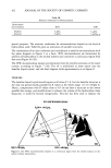
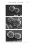
192 JOURNAL OF THE SOCIETY OF COSMETIC CHEMISTS Table I Basal Epidermal ODC Activity and Total Tumor Area in Hairless Mice* Fig. 2 Total ODC activity Total point UVR Continue/ Treatment treatment (nmol CO2/h/ tumor area no. type stop UVR class time (weeks) mg protein) (cm 2) 1 No UVR -- None 12 0.0078 0 2 No UVR -- None 12 0.016 0 3 No UVR -- None 20 0.037 0 4 SSR Continue Chelator 1 + 12 0.392 0.16 sunscreen 5 SSR Stop Vehicle 12 0.602 0.208 6 UVB Stop Vehicle 12 0. 100 0.29 ! 7 SSR Continue Chelator 2 + 12 0.529 0.292 sunscreen 8 SSR Continue Sunscreen !2 0.572 0.355 9 UVB Continue Chelator 3 !2 0.221 0.490 !0 UVB Continue Antioxidant !2 0.680 0.832 ! ! SSR Continue Chelator 2 !2 0.638 0.846 !2 UVB Stop Chelator 3 20 !. !23 0.909 !3 UVB Continue Vehicle !2 2.795 !.045 !4 UVB Stop Vehicle 20 !. 23 ! !. 230 15 SSR Continue Vehicle !2 2. !72 !. 357 !6 UVB Continue Vehicle !2 2.7 ! 3 !. 490 * The order of this list is based on total tumor area and does not necessarily reflect the rank order of photoprotective efficacy. lator 1 (dipyridylamine) plus sunscreen (Point 4) had 88% less total tumor area and 72% lower basal ODC activity compared to the vehicle-treated, similarly irradiated control mice (Point 15). Thus, in hairless mice, elevated levels of epidermal ODC activity, in response to chronic UVR exposure, are associated with visible skin photo- damage assessed by tumor area. Photoprotective agents that inhibited the damaging effects of chronic UVR exposure also inhibited elevation of basal epidermal ODC ac- tivity. Each photoprotective class used in this study is thought to have a different mechanism of photoprotection. Sunscreens protect skin from photodamage by direct absorption of UVR (36). Antioxidants can scavenge the cell-damaging UVR-induced oxygen radicals (29). Iron chelators are photoprotective, presumably by sequestering iron, thereby pre- venting the iron-catalyzed formation of hydroxyl radical from UVR-induced superoxide and hydrogen peroxide (30). Hydroxyl radical is very reactive and can damage a wide range of biological targets. HUMAN BASAL EPIDERMAL ODC ACTIVITY IN SOLAR-EXPOSED VERSUS UNEXPOSED SKIN SITES The next step was to determine if basal epidermal ODC activity is elevated in human skin chronically exposed to solar radiation. To do this, enzyme activity was measured at several different skin sites with varying degrees of solar damage. The results of this study are shown in Table II. No obvious relationship was observed between the basal level of epidermal ODC activity and the level of photodamage. For example, in subjects 1 and 2, skin sites showing very little photodamage (buttock, inner arm) exhibited the highest ODC activity. Conversely, subject 4 showed the highest basal enzyme activity

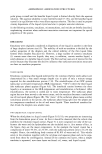
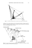
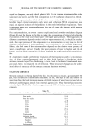
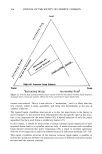
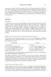
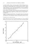
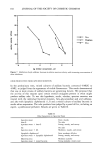
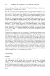
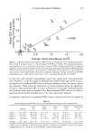
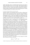
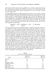
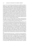
UV-INDUCED SKIN TUMORS 193 "-' 5.0 E2.0 o• 1.0 m E 1 0.0 0.0 12 0 r=0.8 5 10 40 b 1 90 i I I I 0.5 1.0 1.5 2.0 Average Tumor Area/Mouse (cm 2) Figure 2. Correlation between basal epidermal ODC activity and total tumor area in hairless mice (plot of data in Table I). Groups of mice (15-20) were exposed to UVR for 15 weeks, then treated with photopro- tective product for 12 or 20 weeks in the presence (continue UVR) or absence (stop UVR) of continued irradiation. At the end of the treatment period, five mice from each group were killed and basal epidermal ODC activity determined in the non-tumor-involved, UVR-exposed treated dorsal skin. The average total tumor area per mouse was determined from all the animals in each group. Average standard error for ODC determinations was _ 38% and for tumor areas, _ 25%. Data point descriptions are in Table I. in skin sites with extensive photodamage (outer arm, dorsal neck, and preauricular area). Therefore, in the four subjects examined who lacked visible signs of skin cancer, the basal level of epidermal ODC activity did not consistently reflect the extent of prior solar exposure. These results are reminiscent of previous animal studies. No significant increase in basal epidermal ODC activity is observed in chronically irradiated hairless mice lacking visible signs of neoplasia (18). Basal epidermal ODC activity in UVR-ex- posed non-involved skin increased only at the time tumors first appeared. In summary, basal levels of epidermal ODC activity correlate well with the degree of Table II Human Basal Epidermal ODC Activity in Chronically Solar-Exposed and Unexposed Skin Inner Upper Outer Dorsal Preauricular Buttock arm back arm neck area Subject ! 411.6 (0)* 669.3 (0) 16.3 (1.5) 10.1 (3) 24.5 (5) 64.3 (3.5) Subject 2 325.0 (0) 387.8 (0) 149.7 (1) 25.4 (3) 12.0 (3.5) 20.2 (4) Subject 3 10.5 (0) 89.1 (0) 45.3 (2) 53.4 (4) 294.6 (5) 26.5 (4.5) Subject 4 40.6 (0.5) 12.0 (0) 34.3 (2.5) 169.1 (2) 207.6 (3.5) 137.5 (1.5) Average 197 (0.1) 290 (0) 61.4 (1.75) 64.5 (3) 134.6 (4.25) 62.1 (3.75) * ODC activity expressed as pmols CO2/h/mg protein. Numbers in parenthesis represent skin photo- damage grades on a 0-5 scale: 0, no photodamage 5, severe photodamage.
Purchased for the exclusive use of nofirst nolast (unknown)
From: SCC Media Library & Resource Center (library.scconline.org)