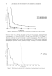
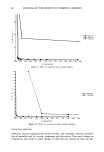
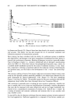
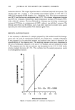
J. Soc. Cosmet. Chem., 42, 71-85 (March/April 1991) Phospholipid liposomes/surfactant interactions as predictors of skin irritation URSULA K. CHARAF and GERALD L. HART, Physical Research, SC Johnson Wax, Racine, WI 53403 (U.K.C.) and Milton Park, U.K. (G.L.H.) Received March 12, 1990. Synopsis The use of large unilamellar liposomes is described as a model membrane system to study surfactant-skin interactions. The relative tendency of surfactants or surfactant blends to form mixed micelles with liposome membrane components determines the aggressivity factor believed to be related to in viva surfactant irritation responses. The potential irritancy ofa surfactant is partly determined by chemical structure, steric factors, and relative polarity, as well as by charge and type of counterions. Single surfactants and surfactant mixtures were investigated and their behavior toward liposomal membranes was used to establish a math- ematical index of surfactant aggressivity. Statistically significant rank correlation was established between this index and in viva scores for the anionic surfactants and blends tested, as well as for some mixed blends, but not with nonionics. The method has distinct advantages over traditional in viva tests. It is expedient, inexpensive, sensitive, objective, and reproducible. Moreover, it does not use live animals or derive its components from tissues or organs. INTRODUCTION Manufacturers of personal care and cosmetic products strive to provide assurances that their products are safe to use. To accomplish this they have relied on conventional in vivo irritancy methods such as the 21-day occlusive patch test (1) and the Draize rabbit eye irritation test (2). These procedures are expensive and time-consuming. They involve monitoring destructive or inflammatory changes in the tissues exposed to surfactants. The readings are subjective and are often unable to discriminate between products with similar mild irritation potentials. Moreover, the Draize eye irritation test and other toxicological procedures involving live animal models have come under public attack for causing suffering. Efforts are being made by very diverse institutions to find alternative approaches (3-5). Liposomes are formed when phospholipids or other suitable amphiphiles are dispersed in an aqueous medium. They aggregate to form vesicular structures consisting of one to multiple lipid bilayers surrounding an aqueous interior. The cell membrane is the most significant example of this lipid bilayer type of structure. The lipid bilayer forms the basic continuous boundary between the interior of the cell and the extracellular space. 71

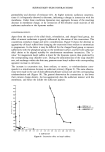
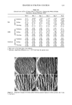
72 JOURNAL OF THE SOCIETY OF COSMETIC CHEMISTS Other membrane components, such as proteins, are embedded in it and depend on the fluidity of the lipid membrane for carrying out many of their functions. For this reason liposomes have been widely used as model membrane systems since their original conception (6,7) to study factors affecting membrane transport, membrane integrity, and disruption. In assessing the damaging effects of extraneous agents on cells, the use of liposomes greatly simplifies dealing with the complexity and variable composition of in vivo systems, while the basic properties of lamellar lipid bilayers common to all cellular membranes are retained. This report describes the development of an in vitro indicator of surfactant irritation on the skin, measuring leakage of a fluorescent marker from phospholipid liposomes ex- posed to surfactant solutions. This method is objective and sensitive and is shown to be capable of indicating the relative irritation potential of a series of anionic surfactants and anionic blends. A mathematical index is developed to facilitate correlation of the data with currently used in vivo indices. It is not expected that the liposome method alone would replace any of the in vivo methods currently in use it may, however, serve as one of a battery of tests that together would give a comprehensive assessment of skin irritancy. The use of liposomes for this particular application is further indicated by findings that correlate skin irritancy directly with the ability of a surfactant system to solubilize skin lipids (8,9). Removal of skin lipids, in turn, leads to a variety of undesirable symptoms, which range from dryness, tightness, scaling, itching, and burning to cracking, in- flammatory changes and, eventually, to a breakdown in the barrier function of the skin (10-12). MATERIALS AND METHODS Egg phosphatidylcholine was purchased from Sigma Chemical Co., St. Louis (Type V-E, 99% in chloroform solution), cholesterol from Nucheck Prep, Elysian, Minnesota, and dicetylphosphate from Aldrich Chemical Co., Milwaukee, Wisconsin. The reference compounds taurodeoxycholate and sodium dodecyl sulfate were purchased from Calbi- ochem, LaJolla, California, and BDH Chemicals Ltd, Poole, U.K., respectively. 5(6)- Carboxy fluorescein (Eastman Kodak) was further purified prior to use (13,14). The surfactants tested were of industrial grade from various sources and were used as sup- plied. PREPARATION OF LIPOSOMES (FIGURE 1) Unilamellar liposomes were produced by the petroleum ether evaporation technique adapted from that described by Schieren et al. (15). Water-jacketed glass chambers resembling micro condensers were custom made by D&H Glassblowing, West Bend, Wisconsin. Phosphatidyl choline, dicetyl phosphate, and cholesterol, in a ratio of 7:2:1 (16,17), were dissolved in chloroform to which a minimal amount of methanol was added. The mixture was then dried under reduced pressure at 45øC for several hours. The dried residue was kept under argon at 4øC until use, when it was dissolved in 35 ml of petroleum ether (Aldrich Chemical Co.) (boiling range: 35-60øC). The solution was then divided evenly into two gas-tight syringes (Hamilton, Reno, Nevada) and
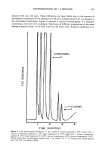
Purchased for the exclusive use of nofirst nolast (unknown)
From: SCC Media Library & Resource Center (library.scconline.org)