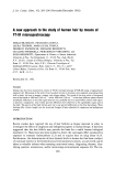
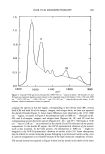
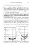
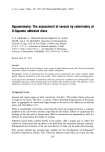
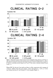
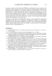
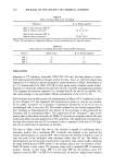
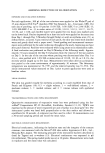
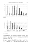
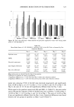
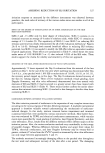
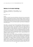
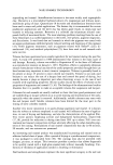
COLORIMETRIC ASSESSMENT OF XEROSIS 299 We also used terephtalate polyethylene sheets (Melinex O, ICI Plastic Division) coated with cyanoacrylate glue (Bison © Super-colle, Perfecta Chemie, Goes, Holland) to re- move surface scales, as previously described (1,4). The cyanoacrylate skin surface biop- sies (CSSB) were stained with a solution of toluidine blue and basic fuchsin (PMS: Polychrome Multiple Stain, Dermatologic Lab Supply, Bluffs, USA). The specimens were then graded (4) accoMing to the following scheme depicted in Figure 2: 0: Normal stratum corneum. 1: Hyperkeratosis of the 1st and 2nd lines and/or of the appendageal orifices. 2: Scales covering less than 30% of the plateaus. 3: Scales covering more than 30%. 4: Diffuse confluent scales. 5: Thick, uneven scales covering the entire surface, obliterating the 1st lines. We also sampled the outer horny layer by means of D-Squame © discs (Cuderm Cor- poration, Dallas, TX). These were applied with a dyanometer under standardized pres- sures of 50, 80, 110, 160, and 210 g/cm 2, respectively, to adjacent sites. These were weighted before and after sampling on a Mettier balance (Zurich, Switzerland) the accuracy of which reaches 0.1 mg. Thus the weight of the stratum corneum sample on each tape was measured as the difference in the weight before and after sampling the skin surface. The samples were then stained for one minute by dropping the staining solution PMS over the surface, followed by gentle rinsing with tap water. The stain solution and rinse water were centrifuged to insure that there was not an appreciable loss of corneo- Figure 2. Examples of CSSB graded 0 (a), 1 (b), 3 (c), and 5 (d).

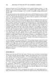
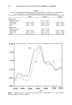
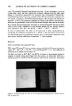
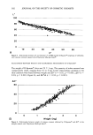
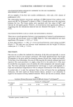
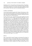
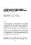
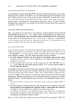
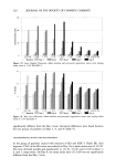
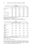
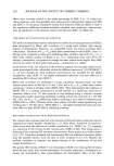
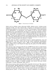
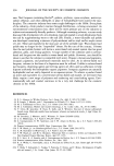
300 JOURNAL OF THE SOCIETY OF COSMETIC CHEMISTS cytes. The stained D-Squame © were placed over a hole, 16 mm in diameter, cut out of a glass slide, which was then placed onto a Minolta reference plate for 0ehite color (Figure 3). Colorimetric parameters were measured with a Chromameter © CR200 (Mi- nolta) following classical operating principles (8,9). We determined the coordinates L*, a*, and b*, according to the CIE International System. We calculated the Chroma C* after (a '2 q- b'2)•/2 this parameter combines the values of red and blue chromaticities, which are the predominant colors of the Polychrome Multiple Stain used in our study. As a control, the same evaluations were done on stained D-Squame © that had not been used to collect scales. Differences in color between samples and our reference material were expressed as AL*, AC*, and AE*ab = [(/•L*) 2 + (/•a*) 2 + (/•b*)2] •/2. A series of measurements was made on the same skin to assess reproducibility by calculating the coefficients of variation (V = SD.M-•). The Chi-square test was used for determining statistical significance between series of evaluations. Regression lines were plotted, and the coefficients of correlation "r" were calculated. RESULTS EFFECT OF PRESSURE UPON D-SQUAME © TAPES PMS stained D-Squame © without stratum corneum yielded the following colorimetric values: L* = 92.2, a* = 1.8, b* = 2.5, and Chroma C* = 3.1. The coefficient of variation on 25 samples was less than 1%. No effect was found on AL*, AC*, and AE*ab in relation to the pressures applied to samples of normal looking skin (Figure 4A). By contrast, on xerotic skin there was a significant positive correlation between pressure and the same colorimetric assessments Figure 3. D-Squame © placed over a hole cut out of a glass slide and deposited onto a Minolta reference plate for white color.
Purchased for the exclusive use of nofirst nolast (unknown)
From: SCC Media Library & Resource Center (library.scconline.org)