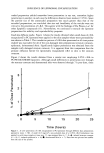
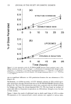
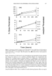
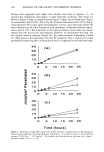
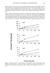
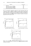
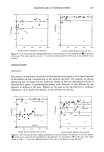
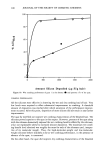
120 JOURNAL OF THE SOCIETY OF COSMETIC CHEMISTS such a change in delivery pattern would be expected to widen the safety margin asso- ciated with topical use. Somewhat awkward for liposome proponents is the lack of a coherent explanation of how liposomes might increase drug concentrations in skin while reducing systemic absorp- tion. The phenomenon implies that liposome components penetrate through the stra- tum corneum and affect drug clearance from the underlying tissues. Yet, careful inves- tigations have failed to show appreciable concentrations of either intact liposomes or of liposome components (phosphatidylcholine or cholesterol) in lower skin layers following topical applications (6,14,18-20), and to our knowledge, no effects of liposomes on cutaneous blood flow have been reported. Thus, the mechanism for the liposome effect remains a mystery, and one is led to examine the methods by which the unexpected results were derived. Most of the evidence for increased drug concentrations in skin following liposomal application has been obtained from skin stripping experiments. This technique, while useful for estimating drug levels in the stratum corneum, does not clearly differentiate between the stratum corneum and the underlying skin layers. We reasoned that a properly controlled technique that measured delivery rates through increasingly thick layers of skin should be able to identify formulations leading to drug accumulation in the lower skin layers. Either the input rate (flux through the stratum corneum) must be higher or the output rate (flux through the whole tissue) must be lower in order for accumulation to occur. With this in mind, we studied the flux of liposomally encap- sulated t-RA vs unencapsulated controls through human stratum corneum, heat- separated human epidermis, and dermatomed human cadaver skin. We studied two liposome systems--a simple soy phosphatidylcholine system and a more complex, four- component system claimed to deliver enzymes through skin (21,22)--and compared their performance against an ethanolic vehicle and a saturated t-RA solution in transcutol/water. We also tested the influence of dosing conditions (dose volume, oc- clusion) on the penetration results and used an inert membrane to identify purely thermodynamic effects. This allowed us to determine whether or not a specific skin/ liposome interaction was taking place. MATERIALS AND METHODS MATERIALS Soya phosphatidylcholine (PC) and phosphatidylethanolamine (PE) were obtained from Avanti Polar Lipids (Alabaster, AL). Cholesteryl hemisuccinate (CHEMS), oleic acid (OA), ot-tocopherol, all trans-retinoic acid (t-RA), [11, 12-3H(N)]-retinoic acid ([3H]t- RA, specific activity 49.30 Ci/mmol), bovine pancreas trypsin, type II-S soybean trypsin inhibitor, N-2-hydroxyethylpiperazine-N'-2-ethanesulfonic acid (HEPES), Dul- becco's phosphate-buffered saline (PBS), and EDTA (disodium salt) were purchased from Sigma (St. Louis, MO). Transcutol (diethylene glycol monoethylether) was obtained from Gattefosse (Elmsford, NY). Tritiated water (specific activity 1.6 ptCi/ml) was obtained from Dupont/NEN (Boston, MA). All other chemicals were of analytical grade.

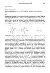
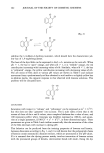
INFLUENCE OF LIPOSOMAL ENCAPSULATION 121 METHODS Preparation and characterization of liposomes Preparation. Liposomes were prepared by a combination of Bangham's film hydration method (23) and the extrusion technique described by Nayar et al. (24). First, t-RA, trace amounts of [3H]t-RA, and the lipids in chloroform solutions were mixed together in the appropriate ratio and 1% of ot-tocopherol (based on total lipid weight) was added as an antioxidant. For PC/t-RA liposomes, the ratio was 16 •mol PC, 4 •mol t-RA, and 50 •Ci [3H]t-RA for the low-dose studies, and 16 •mol PC, 4 •mol t-RA, and 1.2 •Ci [3H]t-RA for the high-dose studies for PC/PE/OA/CHEMS/t-RA liposomes, it was 4 •mol PC, 4 •mol PE, 2 •mol OA, 10 •mol CHEMS, 4 •mol t-RA, and 50 •Ci [3H]t-RA for the low-dose studies, and 4 btmol PC, 4 •mol PE, 2 I-tmol OA, 10 •mol CHEMS, 4 •mol t-RA, and 1.2 •Ci [3H]t-RA for the high-dose studies. The lipid- drug mixture was deposited as a thin film in a round-bottom flask by roto-evaporating the chloroform (Rotavapor RE 111 ©, Buchi, Switzerland) under a gentle nitrogen stream. Vacuum was applied for one hour to ensure total removal of trace solvents. The film was then hydrated at 40øC for one hour with HEPES buffer (20 mM HEPES, 150 mM NaC1, 0.1 mM EDTA). HEPES buffer, pH 7.4, was used for the PC liposomes and HEPES buffer, pH 8.0, for the PC/PE/OA]CHEMS liposomes. After hydration was complete, the preparation was sonicareal for 30 rain (Ultramet III sonic cleaner, Buehler Ltd., Evanston, IL) and five freeze-thaw cycles were performed (5 min, dry ice/40øC waterbath). When necessary, the pH of the PC/PE/OA/CHEMS dispersion was adjusted back to 8.0 with 1 N NaOH. The resulting large multilamellar vesicle dispersion was then transferred into a stainless steel extrusion device (The Extruder TM, Lipex Biomembranes, Vancouver, BC), and unilamellar liposomes were generated by forcing the preparation through two stacked polycarbonate filters (Nuclepore Corp., Pleasanton, CA) of defined pore size (400 nm, 200 nm, 100 nm, or 50 nm in diameter). Nitrogen pressures up to 2800 kPa (400 psi) were used, and all extrusions (ten passes for each preparation) were performed at room temperature. After extrusion was complete, the volume of preparation was adjusted with buffer so that the final suspension contained 0.05% w:v [3H]t-RA. For the low-dose studies, [3H]t-RA specific activity in the final preparation was 12.5 •Ci/•mol, and its radiochemical concentration was 21 •Ci/ml. For the high-dose studies, [3H]t-RA spe- cific activity was 0.3 •Ci/•mol, and its radiochemical concentration was 0.5 •Ci/ml. The final lipid concentration was about 6 mg/ml in both cases. The liposome prepara- tions were stored at 4øC under nitrogen before use. Size determination. We determined the particle size distribution of the extruded liposomal systems by quasi-elastic light scattering (QELS) using a Malvern 4700c submicron particle size analyzer (Malvern Instruments, Southborough, MA) equipped with a 60 mW helium-neon laser at an excitation wavelength of 633 nm. The temperature was set at 25øC and measurements were taken at 90 degrees. In vitro skin permeation studies Preparation of control formulations. For the low-dose studies, we prepared an alcoholic [3H]t-RA formulation (50 •Ci/ml, 12.5 •Ci/•mol) by spiking a 0.05% ethanolic solution of t-RA with trace amounts of radioactive t-RA. Alcohol has been shown to be an efficient delivery vehicle for t-RA (25) and is used in commercial topical preparations
Purchased for the exclusive use of nofirst nolast (unknown)
From: SCC Media Library & Resource Center (library.scconline.org)