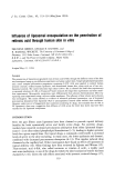
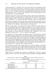
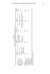
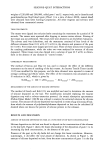
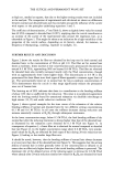
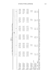
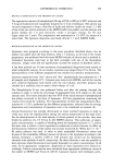
124 JOURNAL OF THE SOCIETY OF COSMETIC CHEMISTS Table II Influence of Liposome Composition and Filter Pore Diameter on Particle Size Distribution Liposome composition Filter pore Distribution mode diameter (nm) (nm) PC No extrusion 1000 100 50 50 26 PC/t-RA a No extrusion 5:1 mole ratio 400 152 200 20 80 b 100 14 70 b 50 10 36 b PC/PE/OA/CHEMS No extrusion 1000 2:2:1:5 mole ratio 400 85 200 48 100 29 50 13 PC/PE/OA/CHEMS/t-RA a No extrusion 1000 2:2:1:5:2 mole ratio 400 85 200 70 100 92 Corresponds to a final t-RA concentration of 0.05% w/v. Bimodal distribution. bution. Extrusion pressures were 3-4 times higher than those of the empty systems. For the PC/t-RA liposomes, extrusion through pores below 400 nm in diameter yielded samples exhibiting bimodal intensity distributions (first mode around 15 nm, second mode in the liposome range). Since it is not possible to achieve a liposome diameter below about 20 nm due to steric constraints, we suspected that these systems also contained miceliar structures. The size distributions of the extruded systems were unchanged after two weeks however, after one week the non-extruded sample had fiocculated. For the PC/PE/OA/CHEMS/t-RA liposome preparations, no miceliar struc- tures were detected, but regardless of the filter used for extrusion, the distribution modes were all about 80 nm. Although a detailed investigation of the effects of t-RA on liposome stability was beyond the scope of our investigation, it seems likely that the explanation may involve an increased propensity to form H-II phases or other non- bilayer phases (30). We concluded that the size distribution of liposomes prepared by the extrusion tech- nique cannot always be linked to the filter pore diameter. Furthermore, incorporation of a lipophilic permeant like t-RA into the formulation can significantly alter liposome structure. Thus, we chose to extrude our liposome preparations through filters with 400-nm pores and to routinely size them prior to use. IN VITRO SKIN PERMEATION STUDIES Efj•ct of liposome size on penetration. The penetration of [3H]t-RA from the PC/PE/OA/ CHEMS/t-RA liposomes in Table II was measured through dermatomed skin (data not shown). The three extruded preparations gave equivalent t-RA penetration. The unex-

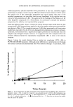
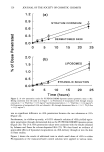
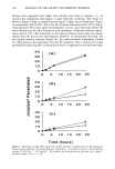
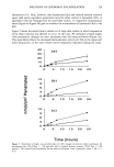
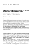
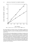
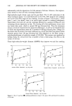
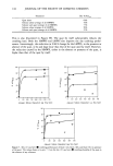
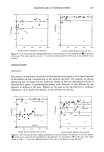
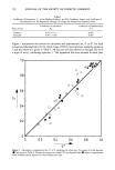
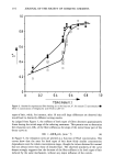
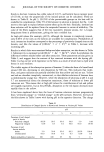
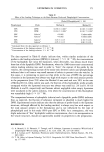
INFLUENCE OF LIPOSOMAL ENCAPSULATION 125 truded preparation yielded somewhat lower penetration in one test, somewhat higher penetration in another in each case the differences observed were modest (25%). Since the particle size of the unextruded preparation was much greater than that of the extruded preparations, we concluded that size and lamellarity of the vesicles were not critical to the penetration of t-RA. This agrees with the findings of Du Piessis et al. for other lipophilic compounds (11). Nevertheless, we continued to extrude the liposome preparations for stability and reproducibility purposes. Small-dose diffusion studies. Figure 1 shows the results obtained when small doses of t-RA encapsulated in PC liposomes were applied to the skin samples whose water permeability was shown in Table I. The cumulative percent of t-RA dose penetrated at all time points studied was equivalent for all of the non-damaged skin preparations (stratum corneum, epidermis, dermatomed skin). Significantly higher penetration was obtained from the samples with damaged stratum corneum. It is apparent from this comparison that the primary diffusion barrier for liposomally encapsulated t-RA in skin is the stratum corneum. Figure 2 shows the results obtained from a similar test employing 0.05% t-RA in PC/PE/OA/CHEMS liposomes. Although small differences in penetration rate through the stratum corneum and dermatomed skin were observed through 7 h post-dose, there 0 L • 0 5 10 15 20 25 Time (hours) Figure 1. In vitro penetration of t-RA encapsulated in PC liposomes through different skin preparations (geometric mean -+ SE). A small (4.8 mg/cm2), non-occluded dose of a 0.05% t-RA formulation was applied to each tissue sample at time zero. O, tape-stripped skin (n = 4) &, isolated stratum corneum (n = 8) /•, isolated epidermis (n = 6) O, dermatomed skin (n = 7). Error bars not pictured are smaller than the size of the symbol.
Purchased for the exclusive use of nofirst nolast (unknown)
From: SCC Media Library & Resource Center (library.scconline.org)