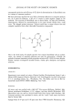
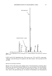
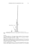
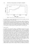
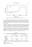
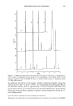
j. Soc. Cosmet. Chem., 45, 193-202 (July/August 1994) Determination of imidurea in cosmetic products by capillary zone electrophoresis and miceliar electrokinetic chromatography GEORGE M. MICHALAKIS, Novo Nordisk Pharmaceutical Industries, Inc., 2231 Powhatan Road, Clayton, NC 27520, and EUGENE F. BARRY, Department of Chemistry, University of Massachusetts, Lowell, MA 01854. Received August 30, 1993. Synopsis Capillary electrophoresis has been employed for the determination of imidurea (imidazolidinyl urea) in cosmetic preparations. When dissolved in water, imidurea consists of 96.6 percent neutral species at a relative standard deviation (RSD) of 0.2 percent and the remaining 3.4 percent is associated with at least twenty anionic species. In addition, the micellar electrokinetic chromatography (MEKC) has been used for the determination of imidurea in the presence of paraben preservatives. The presence of micelles does not affect the imidurea elution profile due to the highly hydrophilic character of the imidurea species observed in this study. The preservatives in several commercial products have been identified and quantitated. INTRODUCTION In the formulation of cosmetic products, preservatives play an important role, since most cosmetics provide an ideal medium for bacterial growth. The parabens have been the most widely used class of preservatives. However, imidurea preservatives became avail- able to cosmetic chemists two decades ago, and their popularity has been steadily increasing. Imidurea is claimed to be nontoxic and nonirritating, with a broad spectrum of antimicrobial activity (1,2). Furthermore, due to the synergistic effect of imidurea with parabens, the two types of preservatives are typically used in combination. Despite the extensive use of imidurea, there are only three analytical procedures ap- pearing in the literature describing imidurea determinations (3-5). Ryder (3) utilized thin-layer chromatography (TLC) and illustrated separations requiring a development time of 50 minutes and an additional 20 minutes at 150øC for visualization. Two spots were observed for the imidurea standard (Rf of 0.27 and 0.35) over a concentration range of 0.1 to 0.6 percent (RSD -- l0 percent, n = 10), and an additional four bands were noted at higher loading experiments. Wilson presented a very extensive TLC procedure focusing on twenty-five preservatives with six indicating reagents where imidurea and propylparaben exhibited Rf values of 0 and 0.6, respectively (4), although detection 193

194 JOURNAL OF THE SOCIETY OF COSMETIC CHEMISTS limits were not reported for either preservative. Sheppard and Wilson (5) described a fluorescence method for formaldehyde-releasing preservatives involving an analytical reaction of liberated formaldehyde with 2,4-pentanedione at 60øC for one hour to generate the fluorophore, and the yield of the reaction was calculated to be 50 percent. The performance of CZE as an analytical technique for the separation of charged species was demonstrated by Mikker and coworkers (6,7), who used 200 }xm i.d. glass and Teflon capillaries, and later by Jorgenson and Lukacs (8-10), who employed 75 }xm i.d. fused silica capillaries. The enormous power of CZE was further extended to neutral compounds with the introduction of MEKC by Terabe and coworkers (11,12). In this communication the use of these modes of capillary electrophoresis is described for the separation of preservatives found in commercial preparations. These techniques yielded excellent separation of complex samples and reproducible quantitative determinations. EXPERIMENTAL APPARATUS Experiments were performed on an Applied Biosystems (San Jose, CA) Model 270A-HT capillary electrophoresis unit with a variable wavelength detector operated at 190 nm and a rise time of 0.50 sec. A HP 3D capillary electrophoresis system (Hewlett-Packard, Palo Alto, CA) was also used for experiments requiring a photodiode array detector (PDA). The fused silica capillary of 50 }xm i.d., 355 }xm o.d. (Polymicro Technologies, Phoenix, AZ) measured 50 cm in length from injector to the detector window. Positive voltage of 30 kV was applied to the injection end of the capillary while the detector end was grounded. The capillary was thermostated at 30øC by forced air convection. Data acquisition was achieved with an IBM personal computer in which SpectraSystem soft- ware system PC1000, version 2. 127 at 30 Hz, was installed. Electropherograms were also displayed with a ChromJet integrator. SAMPLE INJECTION Hydrodynamic sample introduction into the capillary was performed via a controlled vacuum system (@ 5-in Hg) with an injection time of two seconds for most experiments unless otherwise specified. The volume of the sample injected was calculated by the following procedure. The detector was first zeroed with buffer in the capillary, and then the capillary was filled with sample. Buffer was injected into the capillary, and the elapsed time for the detector to reach zero response was noted. For a capillary length of 50 cm, the injection length per second was calculated to be 2.1 mm/sec, corresponding to an injection volume/sec of 0.42 nL/sec. For an injection time interval of two seconds, for example, the injected sample volume was 0.84 nL. CAPILLARY PREPARATION New capillaries were conditioned with 1 N NaOH for at least one hour and subsequently flushed with purified water for a minimum of 30 minutes. Prior to sample injection the capillary was flushed with 0.1 N NaOH for one minute, followed by a water rinse for
Purchased for the exclusive use of nofirst nolast (unknown)
From: SCC Media Library & Resource Center (library.scconline.org)