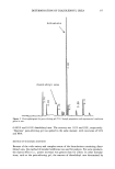
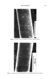
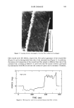
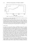
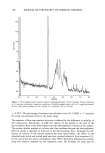
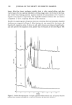
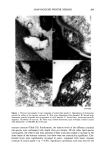
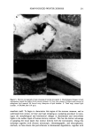
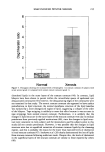
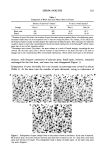
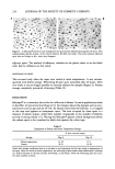
SOAP-INDUCED WINTER XEROSIS 217 such a mechanism would manifest itself in the surface layers of the stratum corneum after only one week of insult to the stratum corneum, which is insufficient time for full stratum corneum turnover, suggesting that the desmosomal and lipid abnormalities we see in the present study are a result of environmental insult to the surface layers of the stratum corneum. It is possible, however, that this mechanism can operate in other more chronic disorders (31,32,42,49). Desmosome retention, however, was also associated with structural abnormalities in stratum corneum lipid lamellae, especially in the outer tape strippings of xerotic skin compared with normal skin. Nevertheless, the gross aberrations in lipid structure were in areas of the tissue where desmosomes were not present and not in the regions where desmosomes were present. The close opposition of corneocytes due to the desmosomal linkages may help to prevent disruption of lipid structure in these parts of the stratum corneum. Although desmosomes largely influence corneocyte cohesion (8), changes in the physical properties of the stratum corneum lipids will also probably influence intercorneocyte cohesion properties and thereby influence desquamation. Changes in the physical properties of stratum corneum lipids are reported to occur in the different layers of the stratum corneum (57). The relationships of these structural changes and skin xerosis remain to be determined. Nevertheless, it has been reported that skin extensi- bility decreases with increasing severity of soap-induced winter xerosis (58). Although we suspect that the diminished elasticity of the stratum corneum arises as a result of increased numbers of desmosomes, changes in lipid structure and reduced natural mois- turizing factors (59) will also contribute. Over three decades ago Kligman (60) suggested that cell cohesion in the stratum corneum was dependent upon an "intercellular cement" that is predicted to become less stable near the surface of the skin or to be degraded by enzymes. We and others (2,12,39,42) have now shown that desmosomal degradation is an important event leading to desquamation. However, we have also provided some morphological evidence for the breakdown of lipid bilayers in the surface layers of the stratum corneum in normal skin. In addition, we also show for the first time changes in the morphology of lipid bilayers and the persistence of desmosomes in severe xerosis. In summary, we have used tape stripping to analyze the roles of lipids and desmosomes in the peripheral layers of the stratum corneum in normal and soap-induced winter xerotic skin. Our results showed that in normal skin there is desmosomal retention close to the surface of the stratum corneum, where their final degradation occurs. Similarily, degradation or disruption of the stratum corneum multiple lipid bilayers apparently occurred at the tissue periphery. The aberrations in soap-induced winter xerosis also appeared to manifest themselves in the upper layers of the stratum corneum, with retention of desmosomes and a collapse of the bilayer structure to give a disorganized lipid matrix of completely different structure to that of normal skin. The resultant interference in the breakdown of the intercorneocyte cohesive forces at the stratum corneum surface appears to account for the scaling that is the major symptom of soap-induced winter xerosis. ACKNOWLEDGMENTS We would like to thank Dr. T. Egelrud, Department of Dermatology, UMEA, Sweden,

218 JOURNAL OF THE SOCIETY OF COSMETIC CHEMISTS for use of his antibody to Desmoglein 1, and Ms. M. DeRosa, Chesebrough Ponds, Connecticut, for the collection of tape strippings. REFERENCES (1) (2) (3) (4) (5) (6) (7) (8) (9) (10) (11) (12) (13) (14) (15) (16) (17) (18) (19) (20) (21) (22) (23) P. M. Elias, Epidermal lipids barrier function and desquamation, J. Invest. Dermatol., 80 (Suppl), 44--49 (1983). S. J. Chapman and A. Walsh, Desmosomes, corneosomes and desquamation. An ultrastructural study of adult pig epidermis, Arch. Dermatol. Res., 282, 304-310 (1990). P.M. Elias, G. K. Menon, S. Grayson, and B. E. Brown, Membrane structural alterations in murine stratum corneum: Relationship to the localisation of polar lipids and phospholipases, J. Invest. Der- matol., 91, 3-10 (1988). H. J. Yardley, Epidermal lipids, Int. J. Cosm. Sci., 9, 13-19 (1987). A. V. Rawlings, I. R. Scott, C. R. Harding, and P. Bowset, Stratum comeurn moisturization at the molecular level, Prog. Dermatol. 28, 1-12 (1994). G. F. Oldland, "Structure of the Skin," In Biochemistry and Physiology of The Skin, L. A. Goldsmith, Eds. (Oxford University Press, 1991), Vol 1, pp. 3-63. M. M. Brysk and J. Miller, Concanavalin A binding glycoproteins in human stratum corneum. J. Invest. Dermatol., 82, 280-282, 1984. S. J. Chapman, A. Walsh, S. M. Jackson, and P. S. Friedmann, Lipids, proteins and corneocyte adhesion, Arch. Dermatol. Res., 283, 167-173 (1991). T. D. Allen and C. S. Potten, Desmosomal form, fate and function in mammalian epidermis. J. Ultrastructural Res., 5, 94-105 (1975). C. J. Skerrow, "Desmosomal Proteins," in Biology of the Integument, 2, Vertebrates, J. Bereiter-Hahn, A. G. Matoltsky, and S. K. Richards, Eds. (Springer Verlag, 1984) pp. 762-787. T. Egelrud and A. Lundstrom, Immunochemical analysis of the distribution of the desmosomal protein, desmoglein 1, in different layers of plantar epidermis, Acta Derm. Venereol. (Stockh)., 69, 470-476 (1989). A. Lundstrom and T. Egelrud, Evidence that cell shedding from plantar stratum corneum in vitro involves endogenous proteolysis of the desmosomal protein, desmoglein I, J. Invest. Dermatol., 94, 216-220 (1990). J. Bartolone, D. Doughty, and T. Egelrud, A non-invasive approach for assessing corneocyte cohesion: Immunochemical detection of desmoglein 1,J. Invest. Dermatol., 96, 596 (1990). T. Egelrud and A. Lundstrom, The dependence of detergent-induced cell dissociation in palmo- plantar stratum corneum on endogenous proteolysis, J. Invest. Dermatol., 95, 456-459 (1990). T. Egelrud, Stratum comeurn chymotryptic enzyme: Evidence of its location in the stratum corneum intercellular space, Eur. J. Dermatol., 2, 50-55 (1992). T. Egelrud, Purification and preliminary characterisation of stratum corneum chymotryptic enzyme: A proteinase that may be involved in desquamation, J. Invest. Dermatol., 10, 200-204 (1993). A. Lundstrom and T. Egelrud, A chymotrypsin-like proteinase that may be involved in desquamation in plantar stratum corneum, Arch. Dermatol. Res., 283, 108-112 (1991). A. Lundstrom and T. Egelrud, Stratum comeurn chymotryptic enzyme: A proteinase which may be generally present in the stratum corneum and with a possible involvement in desquamation, Acta Derm. Venereol. (Stockh)., 71, 471-474 (1991). Y. Suzuki, J. Nonura, J. Hori, J. Koyama, M. Takahashi, and I. Horij, Detection and characteri- sation of endogenous protease associated with desquamation of stratum corneum, Arch. Dermatol. Res., 285, 372-377 (1993). M. M. Brysk, T. Bell, and S. Rajaraman, Sensitivity of desquamin to proteolytic degradation, Pathobiology, 59, 109-112 (1991). S. A. Long, P. W. Wertz, J. S. Strauss, and D. T. Downing, Human stratum corneum polar lipids and desquamation, Arch. Dermatol. Res., 277, 284-287 (1985). A. W. Ranasinghe, P. W. Wertz, D. T. Downing, and I. C. Mackenzie, Lipid composition of cohesive and desquamated corneocytes from mouse ear skin,J. Invest. Dermatol., 86, 187-190 (1985). M. L. Williams, The ichthyosis--pathogenesis and pre-natal diagnosis: A review of recent advances. Pediatr. Dermatol., 1, 1-24 (1983).
Purchased for the exclusive use of nofirst nolast (unknown)
From: SCC Media Library & Resource Center (library.scconline.org)