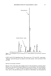
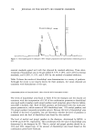
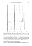
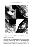
j. Soc. Cosmet. Chem., 45, 221-225 (July/August) An improved procedure for quantitative analysis of sebum production using Sebutape © A. PAGNONI, A. M. KLIGMAN, S. EL GAMMAL, C. POPP, and T. STOUDEMAYER, Foundation for Basic Cutaneous Research, Philadelphia, PA 19103 (A. P., C.P. ), Department of Dermatology, University of Pennsylvania, Philadelphia, PA 19104 (A.M.K. ), Department of Dermatology, Ruhr-University, Bochum 44791, Germany (S.e.G.), and S.K.I.N. Inc., Conshohocken, PA 19428 (T.S.). Received June 3, I994. Synopsis Sebutape © is an adhesive, white tape specifically designed for the collection of sebum. As sebum issues from the orifices, it is trapped in microcavities in the tape, yielding a pore pattern that can be quantified in regard to the number of spots, total area, and size distribution. We have become aware of substantial alterations in pore patterns when Sebutape © is stored for later analysis. These changes relate to enlargement of spots, decrease in their transparency, and increased translucency of the tape itself. We analyzed the evolution of these changes over time and in different storage conditions. We found that placing the Sebutape © on a plastic sheet and storing it in the freezer largely prevented these alterations. Room temperature storage is acceptable only if the samples are evaluated within 24 hours. Analysis immediately after removal yields the least artefacts. Recognizing the importance of changes associated with storage enhances the reliability and accuracy of the Sebutape © method. INTRODUCTION Sebutape © consists of a porous adhesive tape that has been designed to collect sebum on a sebaceous rich area, notably the face. As sebum issues from the follicular orifices, it is trapped within microcavities of the tape and becomes a transparent spot. The area occupied by spots per cm 2 is a measure of sebum production, while the number of spots reflects actively secreting follicles (1-3). Important technological improvements have occurred since the method was first de- scribed. Most recently, image analysis has been used to enable rapid quantitative anal- ysis (2). Usually, the Sebutape © is removed after one hour and placed upon a black card for storage and later analysis. We have become aware of serious changes in the pore patterns after varying periods of storage. In particular, we have noted several changes that can substantially distort the results. These are: a) enlargement of the spots, b) decreased 221

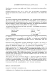
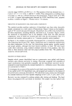
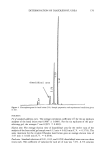
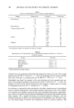
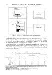
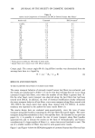
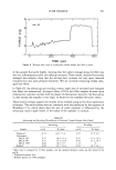
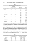
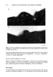
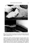
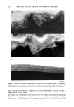
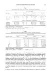
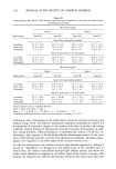
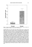
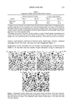
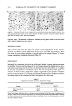
222 JOURNAL OF THE SOCIETY OF COSMETIC CHEMISTS transparency (whitening) of the spots, and c) increased translucency of the white tape, artificially modifying the contrast between the spots and the tape. Our objective was to establish standard conditions that would eliminate the artefacts induced by storage. We were able to prevent these by placing the Sebutape © samples on a plastic sheet rather than a black card and by storing the tapes in a freezer. MATERIALS AND METHODS Sebutape © (CuDerm Corp., Dallas, TX) was applied for one hour to the central fore- head. One sample was placed on the original black storage card (CuDerm Corp., Dallas, TX), while a corresponding one was placed on a transparent plastic report cover (C-Line products, Inc.) that we attached to the original card. Half of the samples was stored at room temperature, while the rest was put in the freezer at - 15øC. In this design, three parameters, enlargement of sebum spots, whitening of spots, and increased translucency of the tape, could be studied under four conditions of storage: a) on a plastic sheet at room temperature, b) on a plastic sheet in the freezer, c) on a black card at room temperature, and d) on a black card in the freezer. Samples were analyzed at times 0 min, 30 rain, 1, 2, 3, 5, 6, 15, and 20 hours, and 1, 2, 3, 4, 7, 14, and 30 days. Sebutape © images from a stereomicroscope (OPMI-1FC, ZEISS, Germany) were trans- ferred to an image analyzer through a black-and-white videocamera (CCD-72, DAGE- MTI, Michigan City, IN). An image analysis program was developed to scan tlae spots (analySIS, Soft-Imaging Software GmbH, Miinster, Germany). The images were pro- cessed with an edge detection filter. The sebum spots were extracted using a gray level scale. Each image was stored on a hard disc. The analysis determined the percentage area covered by sebum spots and their number and mean size. RESULTS INCREASED TRANSLUCENCY OF SEBUTAPE © No increase in translucency was observed on Sebutape © on plastic sheets either at room temperature or in the freezer. By contrast, increased translucency developed, often immediately, when the tapes were fixed on the black card. Storage in the freezer intensified this change, probably because of increased humidity between the tape and the card, while at room temperature this alteration was only minimal. Additionally, in the freezer, translucency gradually increased over time, reaching a plateau after a few days (Table I). ENLARGEMENT OF SPOTS This phenomenon correlated directly with sebum output of individual follicles. Large spots enlarged disproportionately, while small spots underwent marginal changes. At room temperature enlargement reached a peak in 5-15 hours in the freezer the spots slowly but steadily increased, reaching a maximum at seven days. After peaking, the size reached a plateau. The greatest degree of enlargement occurred in the first 30 to 60
Purchased for the exclusive use of nofirst nolast (unknown)
From: SCC Media Library & Resource Center (library.scconline.org)