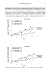
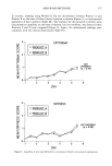
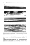
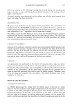
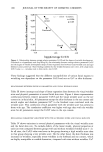
STRATUM CORNEUM LIPIDS 147 Figure 3. Electron microscopy of osmium tetroxide-fixed stratum corneum after (a) 0% and (b,c) 8% extension at 97% relative humidity. Note rupture of desmosome (asterisks). The delipidized tissue has a higher WVTR than the normal tissue without extension. Evidently, extension does not alter the WVTR of the delipidated specimen (delipidated tissue, WVTR = 6.87 -+ 0.05 mg/cm2/h delipidated and extended tissue, WVTR = 6.90 -+ 0.05 g/cm2/h).

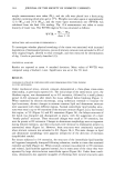
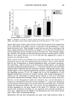
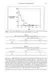
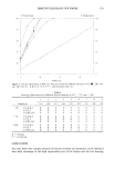
148 JOURNAL OF THE SOCIETY OF COSMETIC CHEMISTS Figure 4. Electron microscopy ruthenium tetroxide-fixed stratum corneum extended to (a) 5% (b) 8%, and (c) 8% at 97% relative humidity and relaxed overnight before fixation. The multiple intercellular lipid lamellae appear marginally disrupted for tissue extended to limited extension (a), but at greater extensions, large intercellular voids are still apparent (asterisks) (b). Note desmosomes appear ruptured and do not reform their correct structure (c). (x 200,000 bar 0.05 DISCUSSION The present study has demonstrated the effects of mechanical extension on the structure of lipids and desmosomes and on the barrier function of the stratum corneum. The observations of mechanical disruption of the intercellular bilayer structures during mechanical extension is a new finding. We were particularly interested in the physio-
Purchased for the exclusive use of nofirst nolast (unknown)
From: SCC Media Library & Resource Center (library.scconline.org)