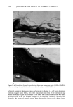
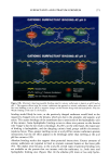
j. Soc. Cosmet. Chem., 48, 219-234 (September/October 1997) Correlation between surfactant-induced ultrastructural changes in epidermis and transepidermal water loss MANOJ MISRA, K. P. ANANTHAPADMANABHAN, KAREN HOYBERG, RICHARD P. GURSKY, SEAN PROWELL, and MICHAEL ARONSON, Unilever Research US, 45 River Road, Edgewater, NJ 07020. Accepted for publication October 31, 1997. Synopsis A comparative analysis of surface and ultrastructural changes in stratum corneum (SC) with transepidermal water loss (TEWL) was conducted to develop a better understanding of the surfactant-induced damage to human skin. Treatments comprised a synthetic hnionic surfactant (A), a composition of soap, glycerin, and petrolaturn (B), a pure soap (C), and water (control). An increase in water loss is shown to correlate with increased perturbation of lipid barrier and damage to multiple layers of corneocytes. Transmission electron microscopy (TEM) revealed that, in general, one to two layers of lipid lamellae enveloped the outer layers of SC throughout the tissue. A significantly larger number of lamellae (approximately six to eight) appeared only in the lower layers. Surface topology, obtained using environmental scanning electron microscopy (ESEM), displayed a normal unperturbed structure. Treatment with A did not result in significant changes in SC. B caused regional variations in corneocytes and an increase in TEWL. Treatment with C exhibited significant variation in TEWL numbers: lipid lamellae were disordered and corneocytes appeared damaged and swollen. Intercorneocyte damage ranged from one to two (for TEWL - 11 g/m 2 hr) to up to six cell layers (for TEWL - 34 g/m 2 hr) of the SC. The presence or absence of the outermost layers of disjunctum did not appear to be critical for water loss, presumably due to a decrease in lipid lamellae. INTRODUCTION Mammalian epidermal cells exist in a state of constant renewal. The process of differ- entiation starts with the embarkation of the basal cells on a well-orchestrated path of terminal differentiation that culminates in the formation of the outermost layer of stratified epithelial cells called stratum corneum. The architecture of the stratum cor- neum is often schematically compared with a brick wall pattern in which the corneocytes (analogous to bricks) lie embedded in a "mortar" of intercellular lipid-rich domains (1). The lipid milieu is highly complex and comprises ceramides, cholesterol, and fatty acids. Cholesterol sulfate, glucosyl ceramides, and phospholipids exist in small amounts (2). Intercellular contact is facilitated by the presence of desmosomes, which form junctions between neighboring cells (3). Depending upon the presence or absence of interlayer desmosomes, the stratum corneum can be further subdivided into compacturn and 219

220 JOURNAL OF THE SOCIETY OF COSMETIC CHEMISTS disjunctum regions, respectively. Degradation of desmosomes is believed to be primarily responsible for the desquamation of the outer corneocyte layers. Some of the important roles of the stratum corneum are to prevent the desiccation of underlying regions, provide moisture retention, and restrict the percutaneous absorption of extraneous substances. The water content of the stratum corneum is dependent upon the quantity of water diffusing into the corneum from underlying layers, its ability to retain water, and the amount of water lost due to evaporation. The barrier property of the stratum corneum (barrier to water loss) is believed to reside in the intercellular lipid lamellae (4) that form a highly ordered, impermeable membranous structure between the corneocytes. An increase in water loss may occur either as a result of decreased ambient humidity or by surfactant-induced cellular perturbations. Dry skin is attributed to defective desquamation, but the mechanisms responsible for surfactant-induced dry skin are not well understood. Although the intercellular lipids are considered responsible for the barrier action of skin, no consensus has emerged regarding the role that the lipids play in dry skin conditions (2). Damage to the ultrastructure of epidermis has been shown to cause decreased water retention properties of the stratum corneum and abnor- mal scaling (5-6). Our current understanding of the influence of surfactants on stratum corneum lipids is not clear, as conflicting evidence exists in the literature. For example, depletion of lipids in surfactant-exposed (7) or solvent-exposed skin (8) is believed to be responsible for increased water loss and for dry skin conditions. On the contrary, Fulmer and Kramer (9) reported that the effect of sodium dodecyl sulfate (SDS) on stratum corneum did not result in lipid depletion. However, significant differences in specific lipid classes were observed. Similarly, lack of damage to lipids was also reported by Fartasch et al. (10), who observed the presence of intact lipid lamellae in the outer layers of surfactant- treated stratum corneum. The present in vitro study attempts to correlate the ultrastructure of the stratum corneum with its water retention properties and the integrity of the barrier after exposure of human cadaver skin to three different surfactant systems. The observed ultrastructural preservation of lipids and corneocytes (using TEM) and surface features (using ESEM) was correlated with TEWL. MATERIALS AND METHODS TREATMENT, WASH, AND TEWL MEASUREMENTS A 4" x 8" piece of abdominal skin from a 31-year-old Caucasian male was mounted (after the removal of the adipose tissue) on an in vitro skin-washing device (11), and the ends of the skin were secured. The temperature of the reservoir was maintained at 37øC using a circulating water bath. The surface of the skin was divided into four equal sections for test treatments using water and three formulations (Table I). Baseline TEWL readings were taken at eight sites in each section. Skin sections were subjected to the following wash procedure: (a) The section was soaked for 15 s under running tap water at 40øC (b) the test bar was wetted under running tap water (40øC) and rotated ten times in the palm (c) the product was applied to the surface of the skin with fingers for 2.0 minutes
Purchased for the exclusive use of nofirst nolast (unknown)
From: SCC Media Library & Resource Center (library.scconline.org)