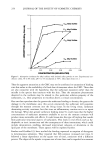
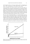
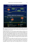
J. Soc. Cosmet. Chem., 48, 235-242 (September/October 1997) Effect of cigarette smoke on skin NEELAM MUIZZUDDIN, KEN MARENUS, P. VALLON, and D. MAES, Estee Lauder Companies, Melville, NY. Accepted for publication October 31, 1997. Synopsis Epidermal barrier quality, skin wrinkling, and skin dryness were compared among groups of active smokers, passive smokers, and non-smokers. Epidermal barrier quality was assessed via transepidermal water loss (TEWL). Skin dryness was measured by digital image analysis of corneocytes that had been removed from skin by tape strippings, and the degree of skin wrinkling was estimated by digitized image analysis of Silflow replicas of the periorbital area. Data obtained from this particular population indicated that active smokers had poorer barrier quality, more wrinkles, and drier skin than non-smokers. Passive smokers presented the same degree of skin barrier damage and skin dryness as active smokers. Sun exposure appeared to play a larger role in the enhancement of skin damage than chronological aging factors, especially in combination with chronic cigarette smoke exposure. Chronic smoke exposure appeared to contribute equally or more than sun exposure to deterioration of barrier quality and the degree of wrinkling. INTRODUCTION Cigarette smoke is still a widespread contaminant in the human environment and has significant health implications for the active smoker (1). Environmental cigarette smoke is a dynamic mixture of sidestream and exhaled mainstream smoke resulting from combustion of tobacco products. It is a complex system, consisting of thousands of compounds, many of which are known carcinogens, cocarcinogens, or tumor promotors (2-4). Smoke also contains numerous free radicals, both in the tar and the gas phases (4,5). Due to their highly reactive nature, these species have potentially damaging effects on the skin surface when interacting with unsaturated intercellular lipids and the membranes of keratinocytes (6,7). Since the frequency of exposure to tobacco smoke is still high and the link between smoking and skin wrinkling is well established (8,9), the effects of such exposure on epidermal barrier function was examined. The results indicate that those exposed to cigarette smoke, either through active or passive smoking, demonstrate significant deficiencies in epidermal barrier function as well as increases in overall surface dryness and wrinkling. MATERIALS AND METHODS The study involved measurements of epidermal barrier function in a total of 100 vol- 235

236 JOURNAL OF THE SOCIETY OF COSMETIC CHEMISTS unteers from the New York, New Jersey, and Pennsylvania area. This population had variable exposure to cigarette smoke. Active smokers were selected on the basis of self-reported histories of heavy smoking (at least one pack a day for over five years). There were forty-five panelists in this sample. Passive smokers were defined as those individu- als who worked or lived with heavy smokers for twenty years, but never smoked themselves. There were thirty panelists in this group. Non-smokers were defined as those who had never smoked and were not exposed to tobacco smoke other than casually such as in a public place. Twenty-five non-smokers were evaluated. All subjects were in normal health, with no evidence of acute or chronic disease includ- ing dermatologic or ophthalmological problems. Subjects exhibiting current sunburn or uneven skin were excluded from the study. The measurement sites were carefully se- lected to be devoid of warts, nevi, moles, sunburn, suntan, scars, and other abnormalities. The volunteers were provided with a mild soap to be used a week prior to the test. They were instructed not to use any skin-care products or moisturizers. The panelists answered a questionnaire to provide a history of sun and cigarette smoke exposure. For sun exposure the panelists graded themselves as having "mild," "moderate," or "severe" sun exposure over past years. Fitzpatrick skin types (10) were determined on the basis of evaluation by a clinical technician. In Part I of the study, the following measurements were obtained: EPIDERMAL BARRIER CONDITION Measurement of epidermal barrier condition in terms of TEWL was made on the cheek area of the face. Measurements were obtained with an evaporimeter (Servomed, Sweden), which allows for determination of water flux just above the surface of the skin (11,12). Prior to water loss measurements, the volunteers remained in the test area at 70øF and a relative humidity of 40% or less, with no air drafts, for 30 minutes to allow for skin equilibration. The panelists were asked to refrain from drinking caffeine-containing beverages prior to coming to the test facility. All measurements were obtained in triplicate from three adjacent sites. SKIN DRYNESS Degree of skin dryness was determined by analyzing the amounts of corneocytes removed on a sticky tape (D-Squame, Cuderm Inc., Dallas, TX) This methodology has been described previously (13). For each subject, measurements were made in quadruplicate. The sticky tapes were analyzed for skin dryness using a digitized image analysis system (I.B.A.S., Zeiss). The D-Squame was placed on a Chroma Pro 45 light box, and its image was obtained via a CCD Panasonic camera. The image was digitized as a 256 gray level image with a resolution of 512 x 512 pixels. The gray value information was extracted from the processed image. The field parameters measured were median and mean stan- dard gray values and its standard deviation. Field area and mean gray values estimate the total number of corneocytes present and the area they occupy. From this information, integrated optical density (IOD) was computed. IOD is indicative of the amount of stratum corneum removed on one sticky tape, an indication of skin surface dryness.
Purchased for the exclusive use of nofirst nolast (unknown)
From: SCC Media Library & Resource Center (library.scconline.org)