SURFACTANT-SKIN INTERACTIONS Table I Principal Constituents of Different Formulations Product Main constituents Formulation A: Isethionate bar Fatty acid isethionate, stearic acid, sodium tallowate, sodium isethionate, coconut fatty acid, sodium sterate, sodium alkyl benzene sulfonate, water, sodium cocoate, fragrance Sodium tallowate/cocoate, glycerin, almond oil, water Sodium cocoate, sodium tallowate, water, fragrance, sodium chloride Formulation B: Glycerin-oil bar Formulation C: Pure-soap bar using a back-and-forth motion, with fingers rewetted after one minute (d) post-wash, the skin surface was rinsed with running tap water (40øC) for 30 seconds (e) steps 2, 3, and'4 were repeated for a total of 15 washes (f) after the final wash, the surface of the skin was patted dry using a paper towel. The apparatus was allowed to equilibrate for one hour. Subsequently, TEWL measurements were taken from the sites used for base- line. The difference between the initial and final values, the delta TEWL value, is presented in Table II. ENVIRONMENTAL SCANNING ELECTRON MICROSCOPY Four 1-cm biopsies from each piece of treated skin were examined in the ElectroScan Model E-3 ESEM. Since no specimen preparation is required for observation using ESEM, the biopsies were placed on a 1-cm aluminum stub for examination in a moist environment. The microscope (equipped with a Peltier cooling stage) was operated at -8øC, and a chamber pressure was maintained at 7.3 Torr. All samples were viewed at an accelerating voltage of 10 kV. TRANSMISSION ELECTRON MICROSCOPY Small pieces of skin (-1 x 2 mm) were dissected from each piece of exposed tissue that corresponded to various TEWL values. Tissue pieces were fixed overnight at 4øC in Table II Transepidermal Water Loss of Tissue Examined by TEM or ESEM A TEWL in g/m 2 hr Specimen Formulation Formulation Formulation no. Water A B C Technique 1 0.37 1.61 2.12 11.53 TEM 2 0.64 2.02 2.30 16.65 3 1.03 2.56 2.76 32.67 4 1.59 2.86 3.98 34.52 5 0.49 1.17 2.54 6.39 ESEM 6 0.56 1.61 3.93 8.99 7 0.66 3.05 4.10 22.34 8 0.92 3.80 4.79 58.35

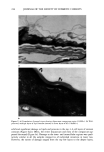
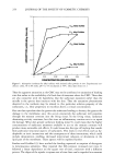
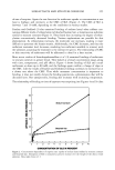
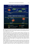
222 JOURNAL OF THE SOCIETY OF COSMETIC CHEMISTS 2.5% glutaraldehyde in 0.1 M sodium cacodylate buffer (pH 7.2). After extensive washing, tissue pieces were post-fixed in either buffered 1% osmium tetroxide (30 min) or 0.5% ruthenium tetroxide (15 min). Samples were rinsed in three changes of buffer before dehydration in a graded concentration series of acetone. Specimens were embed- ded in Spurr resin and incubated at 60øC for two days. Ultrathin sections were cut from the hardened blocks, mounted on carbon-coated copper grids, and post-stained with uranyl acetate and lead citrate. The specimens were examined in a JEOL 1200 EX TEM. RESULTS TEWL TEWL measurements were obtained at eight sites for each of the treatments, and specimens were divided for further structural studies using TEM and ESEM (Table II). The distribution of TEWL measurements was narrow for tissue exposed to water, formulation A, and formulation B. The respective mean (+SD) values of TEWL were 0.78 + 0.4 g/m2hr (water), 2.3 + 0.8 g/m2hr (formulation A), and 3.3 + 0.9 g/m2hr (formulation B). On the other hand, tissue treated with formulation C exhibited the highest TEWL values and showed significant site-to-site variation (24 + 16 g/m2hr). TEM AND ESEM Water-washed control skin. Epidermis, in a water-washed control specimen (Figure la), consists of the inner and outer layers of stratum corneum (compacturn and disjunctum). Disjuncture is characterized by the absence of interlayer desmosomes. In contrast, the corneocytes in successive layers of compactum exhibit the presence of interlayer desmo- somes. The status of desmosomes may differ depending primarily upon the extent of degradation caused by desquamating enzymes. The intercellular space is generally oc- cupied by sheets of lipid lamellae (12), the number and integrity of which vary from region to region. The outermost layers of the disjunctum normally displayed a thin sheet of lipids comprising one to two layers (Figure lb). Departures from this normal outer stratum corneum structure were occasionally observed in those regions of water-washed tissue where stratum corneum appeared either totally or partly devoid of disjunctum (Figure lc). The outer lipid layers, in such regions, displayed robust structures of well-formed multilayers (five or six) that extended around and between desmosomes (Figure ld). In general, the number of lipid lamellae were larger in the lower layers of corneocytes, which also displayed a greater number of interlayer lipid-enveloped des- mosomes. A majority of the tissue, however, displayed intact disjunctum, with very few lipid layers covering the outer corneocytes. Multilamellar lipid layers were seen envel- oping only 20-30% of the surface corneocytes. Structurally, the intercellular proteins and cell envelope appeared normal and remained unaffected by treatment. TEWL values for water-washed tissue pieces did not show significant site-to-site variation. This is confirmed by the uniformity of the structural preservation observed in different pieces of tissue. Even in the 20-30% of the region that displayed damaged disjunctum, the intact lipid multilayers were presumably sufficient to maintain barrier properties. ESEM showed that the surface topography was fairly smooth (Figure 7a), the individual cor-
Purchased for the exclusive use of nofirst nolast (unknown)
From: SCC Media Library & Resource Center (library.scconline.org)