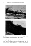
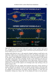
SURFACTANT-SKIN INTERACTIONS 223 neocytes were hardly visible, and very few corneocytes displayed noticeable uplifting (uplifted corneocytes appear brighter with greater contrast). Formulation A-treated skin. The overall ultrastructural morphology of skin exposed to the isethionate bar (formulation A) appeared very similar to that of the water-washed skin. However, in general, fewer cell layers were present in the disjunctum/compactum re- gions (Figure 2a). Thin lamellar sheets or multilayer lipid regions were seen in both formulation A- and water-treated skin. The lipid lamellae in the outer layers were often present as uniform, 4-8 layers thick, well-defined extended regions (Figure 2b) and, as in the water-washed specimens, these regions belonged to the lower layers of stratum comeurn, often in compacturn. The integrity of proteins within the cell and the cell envelope was noticeably unaffected by the treatment. Formulation A-treated skin ex- hibited somewhat higher TEWL values than water-washed skin. This difference may be due to an overall reduction in the number of cell layers and a resultant decrease in the thickness of the corneum. Corneocyte size was unaffected. The surface topography (Fig- ure 7b) was smooth, with a few uplifted corneocytes visible. Formulation B-treated tissue. The glycerin bar (formulation B)-treated specimen was simi- lar to the skin specimen treated with the isethionate bar. The number of cell layers in the disjunctum/compactum region was variable. Although the majority of the tissue displayed the normal morphology of lipid lamallae, evidence of disorder within the lamallae could occasionally be seen (Figure 3a,b). Generally, the organization of cell envelope and proteins was unaltered. In certain cases, however, the tissue displayed significant disruption of proteins and lipids (Figure 4a,b). The intercellular lipids in adjacent outer layers were either absent or displayed disordered structures. Damage to cellular regions was evident in the form of large, globular, low-density regions, which often displayed lipid-like structures (arrows, Figure 4b). Such damage was always con- fined to the outermost layer of the epidermis. Formulation B-washed tissue appeared somewhat more damaged in comparison to either control or to formulation A-treated tissue. This was reflected in terms of somewhat larger TEWL values. ESEM observations also showed the presence of a greater number of uplifted corneocytes compared to control (Figure 7c). Formulation C-treated tissue. The soap bar (formulation C)-treated specimen exhibited maximum ultrastructural damage to cellular proteinaceous material, envelope, and la- meliar lipid structures. The presence of disjunctum could be seen only in about 5% of the treated tissue (Figure 5a). Most of the tissue displayed absence ofdisjunctum and loss of a few layers of compacturn. Lipid structure in the outer layers was often severely damaged. Ordered lamellar structures were replaced by amorphous material. Such re- gions often coexisted with poorly ordered lipid-like structures (arrows, Figure 5b). This could be due to the intercalation of detergent in lipid layers. Intracellular damage to proteins was similar to that described for formulation B except that the damage was much more extensive and severe in the formulation C-washed tissue (almost 25% of the formulation B-washed tissue and 100% of the formulation C-washed tissue displayed intracellular damage). The damaged cells appeared three to four times wider than the control cells (Figures 6a, lc). TEWL measurements showed significant site-to-site variation. The lowest TEWL value for the skin examined using TEM was 11 g/m 2 hr. Damage in this case was confined to the top two layers of corneocytes. Specimens that showed large water loss (-35 g/m 2 hr)

224 JOURNAL OF THE SOCIETY OF COSMETIC CHEMISTS . o Figure 1. (a) Low magnification overview of water-washed tissue displaying the separation between dis- juncture and compactum regions of SC. Disjuncture is characterized by the absence of interlayer desmo- somes (arrowheads) (! ! 000 x). (b) Lipids covering the outermost corneocytes of disjunctum (arrowheads) in control tissue show few multiple layers of ordered lameliar structures (128,000x).
Purchased for the exclusive use of nofirst nolast (unknown)
From: SCC Media Library & Resource Center (library.scconline.org)