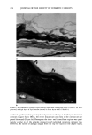
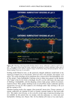
SURFACTANT-SKIN INTERACTIONS 227 Figure 3. (a) Formulation B-treated tissue showing desmosomes (arrowheads) and disjunctum-compactum region of the SC (24,000x). (b) Multiple layers of lipid lamellae (arrows) surrounding the outer layers of SC (128,000x). depending upon TEWL. ESEM micrographs showed that the surface of formulation C-treated skin resembled the surface features of dry skin, i.e. a large number of corneo- cytes displayed uplifted morphology (marked regions, Figure 7d). DISCUSSION This study provided an opportunity to examine the correlation between surfactant-

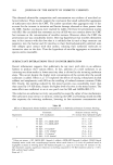
228 JOURNAL OF THE SOCIETY OF COSMETIC CHEMISTS Figure 4. (a) Damage to outer layers of corneocytes (arrows) was seen in 25% of the formulation B-treated tissue (24,000x). (b) A higher magnification view of the damaged region showing fingerprint (arrows) of lipid-like regions surrounding the outer corneocytes (128,000x). induced structural changes in epidermis with TEWL. Different surfactant treatments resulted in variable degrees of lipid and corneocyte preservation. Most of the water- washed tissue (control) displayed intact disjunctum comprising few layers (one to two) of lipid covering the outer corneocytes. In contrast, isolated regions that lacked most of
Purchased for the exclusive use of nofirst nolast (unknown)
From: SCC Media Library & Resource Center (library.scconline.org)