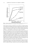
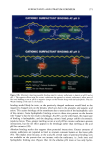
20 MHz B-SCANNING 249 201-255 pixels 900 800 - 700 - 600 - 500 - 400 - 300 - 200 - IO0 baseline rem 15' 30' 1 h 2h 3h • petrolatum • moisturizer 1 • moisturizer2 Figure 4. Image analysis on echographic pictures. Variation of the extension of areas reflecting within the 201-255 interval in the lower dermis. *Significant in respect to baseline values. corneum by two different mechanisms, the first by delivering their water to the skin, and the second by occlusion (9). In fact, it has been demonstrated that lipids contained in petrolatum penetrate to all levels of the stratum corneum, replacing intercellular bilayers, supporting the concept that petrolatum and other moisturizers do not simply form an inert, epicutaneus, oc- clusive membrane, but actively contribute to the hydration process and barrier recovery (15). In our study, too, after removal of the moisturizers, TEWL was higher, owing to evaporation of the water included in the test substances that penetrated into the stratum corneum during the application period. At the same time, capacitance values rose both for the two test formulations and for petrolatum. Contrary to the findings of Marti- Mestres et al. (16), we did not observe a decrease in TEWL after application of petro- latum. Instead, a slight increase in TEWL was assessable even at petrolatum test areas. This different observation may be due to the different experimental procedure employed by these authors, whereby the residues of petrolatum, forming an occlusive membrane, were not removed from the skin before performing measurements. Echographically, dermal modifications induced by topically applied substances causing edema or inflammation are appreciated as an increase in the extension of hypo-refiecting areas (17-20). Occlusion exerted by an empty Finn-Chamber on the skin induces a slight edema that is assessable by ultrasound (21). In this study, image analysis on echographic

250 JOURNAL OF THE SOCIETY OF COSMETIC CHEMISTS pictures revealed a decrease in the echogenicity of the dermis, which was significant for all three products for echographic values assessing deep edema (201-255 pixel values). We can therefore presume that external factors influencing the skin barrier may induce modifications in dermal blood flow causing edema, which is particularly pronounced in the lower dermis. When evaluating an ultrasound scan of the skin, a strong band-like echo is seen at the boundary between the coupling medium and the skin, the so-called entry echo. Some authors equate this structure with the epidermis (22-25), whereas others regard it as a result of the impedence jump at the border between the epidermis and the coupling medium (26). The evaluation of epidermal changes by image analysis procedures has enabled the description of modifications of the reflectivity of the entrance echo, which vary according to the different irritation modalities of the test substances. Detergents, such as sodium lauryl sulfate, induce a decrease in the extension of the superficial hyper-refiecting band, which is invertly related to TEWL values (18, 19). On the contrary, nonanoic acid- induced irritation appears with an increase in the epidermal reflectivity (20). By applying moisturizers and petrolatum, we observed a reduction of superficial 201- 255 pixel areas, whose extension was inversely related to capacitance values. Thus, echographically, the higher the hydration state of the stratum corneum the lower the impedence jump and consequently the higher the attenuation of the epidermal band. This confirms previous observations on an analogous decrease in epidermal reflectivity, associated with an increase in capacitance values and TEWL, when saline solution was applied to the skin under occlusion (27). In conclusion, we suggest that data deriving from image analysis of 20 MHz B-scanning could be used as a support for the findings of other instrumental techniques in the assessment of skin hydration. REFERENCES (1) J. K. Prall, R. F. Theiler, P.A. Bowser, and M. Walsh, The effectiveness of cosmetic products in alleviating a range of skin dryness conditions as determined by clinical and instrumental techniques, Int. J. Cosmet. Sci., 8, 159-174 (1986). (2) R.E. Dunlap, Clinical evaluation of a highly effective hand and body lotion, Curr. Ther. Res., 35, 72-77 (1984). (3) T. H. Cook and T. J. Craft, Topographics of dry skin, non-dry skin, and cosmetically treated dry skin as quantified by skin profilometry, J. Soc. Cosmet. Chem., 36, 143-152 (1985). (4) J. Serup, A. Winther, and C. W. Blichmann, Effects of repeated application of a moisturizer, Acta Derre. Venereol. (Stockh.), 69, 457-459 (1989). (5) S. Nicholls, C. S. King, and R. Marks, Short term effects of emollients and a bath oil on the stratum corneum, J. Soc. Cosmet. Chem., 29, 617-624 (1978). (6) D. Wilson, E. Berardesca, and H. I. Maibach, In vivo transepidermal water loss and skin surface hydration in assessment of moisturization and soap effects, Int. J. Cosmet. Sci., 10, 201-211 (1988). (7) C. W. Blichman, J. Serup, and A. Winther, Effects of single application of a moisturizer: Evaporation of emulsion water, skin surface temperature, electrical conductance, electrical capacitance, and skin surface (emulsion) lipids, Acta Derre. Venereol. (Stockh.), 69, 327-330 (1989). (8) M. Lod•n and M. Lindberg, The influence of a single application of different moisturizers on the skin capacitance, Acta Derre. Venereol. (Stockh.), 71, 79-82 (1991). (9) M. Lod•n, The increase in skin hydration after application of emollients with different amounts of lipids, Acta Derre. Venereol. (Stockh.), 72, 327-330 (1992).
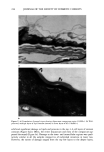
Purchased for the exclusive use of nofirst nolast (unknown)
From: SCC Media Library & Resource Center (library.scconline.org)