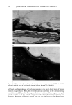
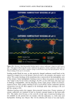
232 JOURNAL OF THE SOCIETY OF COSMETIC CHEMISTS damage was reflected in high TEWL values. The nature of structural damage was similar in all specimens irrespective of individual variations in TEWL. The fact that only the upper layers appeared damaged in tissue displaying lower TEWL numbers (11 g/m 2 hr) in contrast to four to six affected layers for high TEWL numbers (35 g/m 2 hr) indicates a direct relationship between the extent of damage and water loss. TEWL is generally considered to be a passive diffusion phenomenon. Assuming Fick's law of diffusion as a first approximation, diffusion of water could be considered inversely proportional to the distance or the total thickness of the corneum. However, water loss through the surface layers of corneocytes is not only inversely proportional to the thickness of the epidermis but also depends upon the resistance to its migration pathway provided by the barrier lipids. Both parameters (epidermal thickness and lipids) were affected by different surfactant treatments. Although the epidermis often displayed structural variability even in water-treated tissue, the decrease in corneum thickness was not significant enough to affect TEWL, as was evident by the uniformity of TEWL measurements. Also, in water-treated tissue the lipids were well preserved in the lower layers of the stratum corneum, providing an effective barrier against water loss. Treat- ment with the isethionate bar resulted in an increase in the frequency of occurrence of intact multilamellar lipid sheets covering the upper corneocyte layers. This suggests that the treatment with formulation A resulted in a reduction (compared to control) in the outer layers of disjunctum and consequently caused a slight increase in TEWL. The water loss, however, remained limited due to the barrier offered by the surface- contouring lameliar lipids. Glycerin bar-treated tissue displayed features similar to those described for the isethionate bar. However, the outer lipids displayed damaged mor- phology, i.e., non-lamellar, amorphous material in about 25% of the surface regions. It is tempting to suggest that the modest increase in TEWL in glycerin bar-treated tissue could be ascribed to damaged lipids in the outer layers of surface corneocytes, but the relationship is only directional at best. Significant dependence of TEWL on the thinning of epidermis and disrupted lipid structures was evident in soap bar-treated tissue. The disjunctum was compromised in almost 90% of the tissue. The lipid barrier underlying the disjunctum was severely affected and appeared as an amorphous non-lamellar struc- ture. In tissue displaying 30 g/m 2 hr of water loss, the stratum corneum was punc- tuated with regions in which almost all structure above the viable cell layers was disrupted (Fig. 6c). The soap bar-treated tissue revealed a decrease in the number of cell layers forming the stratum corneum, lipid loss, and significant swelling of the corneocytes. Some of the corneocytes were four times wider in projection compared to the normal corneocytes. A surfactant-induced increase in surface area and swelling is known to depend on the surfactant type (13). Increased permeability of the cell membrane and consequent in- crease in the intracellular binding of water molecules may be responsible for the observed increase in the projected width of damaged corneocytes. An advantage of using ESEM for structural studies is that the specimen can be observed in its native state without preparation artifacts. However, the interpretation of the ESEM micrographs requires a careful analysis. Normal, undamaged skin in ESEM appears as a smooth surface. In damaged skin, the structural changes to corneocytes appear as brighter regions. This could be attributed to the uplifting of cells, which causes changes in their geometric disposition. Peripheries of uplifted corneocytes appear brighter than surrounding regions. Such fine changes are often difficult to detect because the skin

SURFACTANT-SKIN INTERACTIONS 233 surface is rarely flat and signal variations from neighboring (-100 pm) regions may also contribute to significant intensity variations in the image. Therefore, extraction of fine structural changes (in individual comeocytes) from gross intensity variations requires a careful analysis of the micrographs. The accuracy of TEWL measurement is dependent upon the amount of water loss. Measurements are highly reproducible for low-water-loss conditions such as those that exist in normal unperturbed skin. If the water loss is about 20 g/m 2 hr, the measurement error could be about 10%. However, when losses approach 80 g/m2hr, the error could exceed 50% (14). TEWL in surfactant-treated tissue may be as high as 60 g/m • hr, as was seen for soap bar-treated tissue. However, the exact TEWL value is not significant for the current discussion, as the dependence of structural damage on TEWL values was fairly consistent. The high degree of water loss encountered in soap bar-treated tissue may be due to the combined effect of damage incurred to lipids and to the intracellular environment. This could also affect the water-retaining mechanisms involving the role of molecules re- sponsible for providing natural moisturizing factor (15). Leaching of such molecules from surfactant-damaged corneocytes may aggravate dry skin condition. This study showed that the presence or absence of outer layers of disjunctum, which have minimal surrounding lipid, is not critical for water loss. Water loss depended upon the preservation and integrity of lipids and proteins belonging to the lower layers of cor- neocytes. No severe water loss was observed in cases where the lipids in compactum appeared ordered and structured. Inordinately high water loss (for the soap bar) was due to damage that extended from the surface to multiple strata of underlying lipids and comeocytes. REFERENCES (1) P.M. Elias, Epidermal lipids: Barrier function and desquamation,J. Invest. DermatoL, 80 (Suppl), 44- 49 (1983). (2) A. V. Rawlings, A. Watkinson, J. Rogers, A. Mayo, J. Hope, and I. R. Scott, Abnormalities in stratum corneum structure, lipid composition, and desmosome degradation in soap-induced winter xerosis, J. Soc. Cosmet. Chem., 45, 203-220 (1994). (3) S.J. Chapman and A. Walsh, Desmosomes, corneosomes and desquamation. An ultrastructural study of adult pig epidemis, Arch. Dermatol. Res., 282, 304-310 (1990). (4) P. Bowsen and R.J. White, Isolation, barrier properties and lipid analysis of stratum compacturn, a discrete region of the stratum corneum, Br. J. Dermatol., 112, 1-14 (1985). (5) G. Imokawa, K. Sumura, and M. Katsumi, Study on skin roughness caused by surfactants. I. A new method in vivo for evaluation of skin roughness,J. Am. Oil Chem. Soc., 8, 92-108 (1975). (6) G. Imokawa, Evaluation for alteration of the stratum corneum, J. Jpn. Cosmet. $ci. $oc., 8, 92-108 (1984). (7) G. Imokawa, S. Akasaki, Y. Minematsu, and M. Kawai, Importance of intercellular lipids in water retention properties of the stratum comeum: Induction and recovery study of surfactant dry skin, Arch. Dermatol. Res, 281, 45-51 (1989). (8) G. Imokawa, S. Akasaki, M. Hattori, and N. Yoshizuka, Selective recovery of deranged water-holding properties of the stratum corneum lipids,J. Invest. Dermatol., 187, 758-761 (1986). (9) A. W. Fulmer and G. J. Kramer, Stratum corneum lipid abnormalities in surfactant induced dry skin, J. Invest. Dermatol., 86, 598-602 (1986). (10) M. Fartasch, T.L. Diegpan, O. P. Hornstein, Morphological changes of epidermal lipid layers of stratum corneum in sodium lauryl sulfate induced dry skin, J. Invest. Dermatol., 96, 617A (1991). (11) K. P. Ananthapadmanabhan, S. Prowell, K. Hoyberg, M. Misra, S. Spaltro, S. Mukherjee, and M.
Purchased for the exclusive use of nofirst nolast (unknown)
From: SCC Media Library & Resource Center (library.scconline.org)