50 JOURNAL OF COSMETIC SCIENCE POLYMER-STABILIZED LIPOSOMES: PREPARATION, CHARACTERIZATION AND APPLICATIONS Francoise M. Winnik, Ph.D. McMaster University, Hamilton, ON L8S 4M1 Canada Introduction Liposomes are spherical vesicles consisting of one or several bilayers of amphiphilic molecules surrounding an inner aqueous pool. This morphology enables the entrapment of lipophilic materials in the bilayer and of hydrophilic compounds in the aqueous compartment. This ability to transport substances soluble in either oil or water makes liposomes extremely attractive in cosmetics applications. • Other advantages offered by liposomes are their high affinity with biological membranes and their perceived ability to enhance the natural hydrating functions of skin'- and hair. Traditionally, the liposome membrane constituents were selected among natural phospholipids, such as lecithin (phosphatidylcholine), phosphatidylserine, and phosphatidylethanolamine. Unlike other tissues, the stratum comeurn is devoid of phospholipids) Therefore, the membrane of liposomes ideal for topical applications should be composed of amphiphiles similar to skin's lipid lamellae. Natural derivatives, such as ceramides and cerebrosides, when mixed with cholesterol and palmitic acid were shown to form liposomes. 4 More recently, it was reported that stable liposomes can be prepared from a large number of synthetic lipids. 5 These vesicles, known as non-phospholipid liposomes (NPL's), are obtained from a mixture of three types of lipids: (a) a main amphiphile, such as n- octadecyldiethylene oxide, (b) a modulator, usually cholesterol, and (c) an ionogen, such as dioctadecyldimethylammonimn bromide (DDAB) in the case of cationic vesicles or dioctadecyl phosphate (DP) in the case of anionic NPL's. A major shortcoming of liposomes as delivery agents in cosmetics formulations is the fragility of the bilayer membrane, especially in the presence of surfactants. A promising method to strengthen the liposome is to anchor or ligate polymers to the lipid bilayer. In addition, the outer surface of liposomes can be endowed with specific properties by further modifications, such as the attachment of polymers carrying specific receptors or responsive to changes in temperature, pH, or ionic strength. 6 The design and properties of complexes between non-phospholipid liposomes and polymers, such as Quatrisoft LM200 and its fluorescently-labeled derivatives, will be discussed. Techniques used to monitor the properties of liposomes and polymer/liposome complexes will be presented. They include fluorescence spectroscopy, dynamic light scattering, centrifugation assays and gel permeation chromatography. Materials and Methods Cholesterol, n-octadecyldiethylene oxide (EO2C•sH37), and dicetylphosphate (DCP) were purchased t¾om Aldrich Chemicals. Quatrisoft LM200 and (hydroxyethyl)cellulose (HEC, approximate MW 400 000) were gifts of Amerchol Inc. Pyrene-labeled Quatrisoft LM200 (LM200-Py) and HEC-N • PyC•2 were prepared as described previously. 7 It contains 2.9 x 10 -5 mol Py g-• of polymer or on average 1 Py per 190 glucose units. Instrumentation. Fluorescence spectra were recorded on a SPEX Industries Fluorolog 212 spectro•neter equipped with a GRAMS/32 data analysis system. UV spectra were recorded with a Hewlett Packard 8452 photodiode array spectrometer. Dynamic light scattering measurements were performed with a Brookhaven Instrument Corporation Particle Sizer Model BI-90. Liposomes were prepared by extrusion using a Lipofast extruder (Avestin, Canada) fitted with polycarbonate filters (200 nm pore size) obtained from Avestin. Liposome preparation. A solution in chloroform of EO2CtaH37, cholesterol, and DCP (75/20/5 w/w) was poured into glass vials. The solvent was evaporated under a stream of nitrogen. It was dried under high vacuum for at least 2 hr. The dry lipid film was hydrated with aqueous NaCI (I mmol) to give a 20 g L • lipid suspension. The lipid suspension was subjected to extrusion at 65 øC. Liposome/polymer mixtures were prepared by addition of an extruded liposome suspension (2.7 mL) to polymer solutions (0.3 ink) of various concentrations. The mixtures were allowed to equilibrate for at least 2 hr prior to spectroscopic measurements.

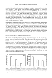
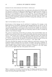
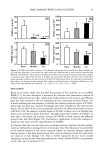
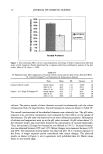
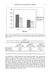
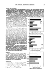
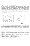
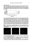
1999 ANNUAL SCIENTIFIC MEETING 51 Results and discussion The interactions of Pyrene-labeled Quatrisoft-LM200 and negatively-charged non-phospholipid liposomes have been investigated by fluorescence spectroscopy and dynamic light scattering, following protocols developed in our previous studies of liposome/polymer systems. 8 The fluorescence assay relies on the relative intensities of pyrene excimer and monomer emissions, 9 as the polymer hydrophobic substituents are inserted within the liposome bilayer. The effect is illustrated in Figure l, which presents the emission spectra of Quatrisofi LM200Py and water and in the pre.sence of negative NPL's, and in Figure 2, where are plotted the changes in the ratio IE/IM determined from emission spectra of Quatrisoft LM200 Py of increasing concentration in the presence of liposomes. The excimer emission all but disappears for polymer solutions of concentrations lower than 0.008 g L -•. This concentration corresponds to the point of saturation of the NPL surface. Above this concentration polymer-coated liposomes and polymer micelies coexist in solution, as indicated also by dynamic light scattering experiments. 4000 m 3000 • 2500 • 2000 m 1500 •O 1000 U. 500 0 350 LM200-Py + NPL 400 450 500 550 600 Wavelength (nm) Figure 1 1 0.8 o.6 0.4 0,2 0.3. 5 LM200Py in water + LM . + NPI -3 -2.5 -2 -1.5 -1 Log[HEC-PyN+C•2] (g L 'l) Figure 2 -0.5 Conclusion The study demonstrate that hydrophobically-modified cationic cellulose ethers interact eftkctively with negatively-charged non-phospholipid liposomes via hydrophobic interactions as well as via hydrogen bonding between the cellulose ether hydroxyl groups and the head groups of the membrane. Potential applications of these complexes as delivery vehicles in cosmetics applications will be discussed. References I See for example: Burnmeister, F. Bennet, S. Brooks, G. Costa. & Toil, 1996, I 11,49 and references therein 2 Suziki K Costa. & Toil. 1990, 95, 5. 3 Scheuplein, R. in The Physiology and Pathophysiology of the Skin, A. Jarrett, Ed. Academic Press, New York NY, pp 1669 (1978) 4 Egbaria, K. Ramachandran, C. Weiner, N. Skin Pharmacol. 1990, 3, 21. 5 Philipot, J. R. Milhaud, P. G. Puyal, C. O. Wallach, D. F. H. In Liposomes as Tools in Basic Research and Industry Philipot, J. R., Schubert, F., Eds. CRC Press: Boca Raton, FL, 1995 Chapter 3. 6 Polozova, A. Winnik, F. M. Langmuir 1999, 15, 4224 and references therein. 7 Winnik, F. M. Regismond, S. T. A. Goddard, E. D. Langmuir 1997, 13, 1 l 1. 8 Ringsdoff, H. Simon, J. Winnik, F. In Colloid-Polymer Interactions ACS Symposium Series, Dubin, P.. Tong, P. Eds. American Chemical Society, Washington, DC, 1993 216. 9 Winnik, F. M. Chem. Rev. 1993, 93. 587.
Purchased for the exclusive use of nofirst nolast (unknown)
From: SCC Media Library & Resource Center (library.scconline.org)