368 JOURNAL OF COSMETIC SCIENCE the state of dissociation of the drug (5-7). To our knowledge, there are no reports in the literature on variations in nail permeability as a function of different sources of nails. The effect of environmental factors such as pH and temperature on intrinsic nail permeability properties also remains to be investigated. The objectives of our research were to (a) develop a reproducible technique to measure nail permeation in vitro, (b) study variations in nail permeability as a function of differing source (between donors, toenails vs fingernails), and (c) study the effect of pH and temperature on nail permeation. In our experiments, tritiated water was chosen as a marker molecule, and its permeation through nails was monitored as a function of various environmental and physical factors. Water was expected to be non-destructive to the nail and is a good marker because of its small size. It was first necessary to develop a suitable technique to reproducibly measure nail permeation in vitro. For this purpose, specialized diffusion cells that had been adapted to hold the human nail were used, and water permeation through the nails was repeatedly monitored. EXPERIMENTAL MATERIALS Tritiated water (3H20) (specific activity of 1 l•Ci/mg) was obtained from NEN TM Life Sciences Products (Boston, MA). Hydroxyethyl cellulose (HEC, Natrosol ©, Aqualon Co., Wilmington, DE) and polyethylene glycol-20-oleyl ether (PEG-20-oleyl ether, Croda, Inc., Parsippany, NJ) were used as received. All other laboratory chemicals (ACS grade or better), including Scintiverse I, were obtained from Fisher Scientific (Springfield, NJ) and used as received. Cadaver toenails or fingernails were obtained (frozen) from tissue banks [International Institute for the Advancement of Medicine (IIAM), Scranton, PA Advanced Biosurfaces Inc., Minnetonka, MN]. DEVELOPMENT OF METHODOLOGY FOR IN VITRO NAIL PERMEATION STUDIES Preparation of nails. Human toenails/fingernails were obtained (frozen) from tissue banks based on a protocol for size, patient history, etc. Of all the toenails, only the great toenail was large enough to fit in the diffusion assembly however, all the fingernails (thumbnail and other fingernails) were suitable for use. The nails were thawed at room temperature for one hour, and the adhering skin and tissue were removed with a pair of scissors and a scalpel. The nails were cleaned by rinsing them in a mild detergent solution (1%), followed by two rinses in aleionized water (DI water). The thickness of the nails was measured with a micrometer, and the nails were immersed in 10 ml of DI water for 24 hours to allow complete hydration just prior to use in permeation experiments. Nail permeation cells. Franz-type diffusion cells (Crown Glass Co., Somerville, NJ), spe- cially designed to hold the human nail were used (Figure 1). In this cell, the donor compartment (Teflon) had a two-part construction to hold the nail. The lower half of the donor compartment screwed on to the glass receptor chamber, the nail was placed above this, and the upper half of the donor compartment was placed above the nail. The two parts of the donor compartment were clamped together to hold the nail in place between

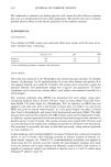
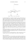
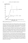
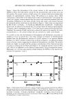
NAIL PERMEATION 369 Donor Compartment (Teflon) Receptor Compartment (Glass) .i Donor Formulation Applied Through Opening Nail Receptor Solution • Water In Water Out Stirrer Bar MagnetStirreSwitchStirrer Figure 1. Schematic of the diffusion cell used for in vitro permeation studies. them. Also, each part of the donor compartment was curved to accommodate the natural curvature of the nail, thus ensuring a good fit. The area of the cell available for perme- ation was 0.2749 cm 2. A circulating water bath maintained the temperature of the receptor compartment at 37øC. Magnetic stirrer bars were used to ensure receptor uniformity throughout an experiment. Donor formulations JCr water permeation studies. Aqueous gels were prepared with 1.5% HEC and spiked with a small quantity of 3H20 so as to obtain a ratio of hot:cold drug of 1:1000. The gels were agitated with a vortex mixer (Vortex Genie, Fisher Scientific, Springfield, NJ) and allowed to stand at room temperature for 24 hours to ensure homogenous distribution of the label. Prior to use, three 10-pl aliquots of the gel were analyzed by liquid scintillation counting (LSC) to validate uniformity of spiking. Permeation studies. To establish and validate a reproducible technique to study nail per- meation in vitro, water permeation studies were run with six replicates using excised, human toenails. The water permeation studies were repeated three times on the same nails to assess the reproducibility of the method. Hydrated, trimmed nails were clamped in the diffusion cells, and the receptor com- partment was filled with the receptor fluid (water containing 0.5% PEG-20-oleyl ether, a wetting agent to facilitate removal of air bubbles and not expected to affect the nail at such a low concentration). The cells were tilted and gently tapped to remove any entrapped air bubbles. A temperature of 37øC and constant stirring was maintained throughout the study. The cells were left uncovered for one hour prior to application of
Purchased for the exclusive use of nofirst nolast (unknown)
From: SCC Media Library & Resource Center (library.scconline.org)