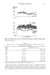
GLYCOLIC ACID IN SKIN CREAMS 349 REFERENCES (1) E.J. Van Scott and R.J. Yu, Control of keratinization with alpha-hydroxy acids and related com- pounds, Arch. Dermatol., 110, 586-590 (1974). (2) M. Dahl and A. Dahl, 12% Lactate lotion for the treatment of xerosis: A double-blind clinical evaluation, Arch. DermatoL, 119, 27 (1983). (3) E.J. Van Scott and R.J. Yu, Alpha-hydroxy acids: Therapeutic potentials, Can. J. Dermatol., 1(5), 108-112 (1989). (4) M. L. Elson, The utilization of glycolic acid in photo-aging, Cosmet. DermatoL, 5, 12-15 (1992). (5) E. M. Jackson, Update on AHA-containing products, Cosmet. Dermatol., 7, 29-30 (1994). (6) CIR, Final report on the safety assessment of glycolic acid, ammonium, calcium, potassium, and sodium glycolams, methyl, ethyl, propyl, and butyl glycolams, and lactic acid, ammonium, calcium, potassium, sodium, and TEA-lactates, methyl, ethyl, isopropyl, and butyl lactates, and lauryl, myri- styl, and cetyl lactates, Int. •. ToxicoL, 17(Suppl. 1), 1-242 (1998). (7) E. Christophers and A.M. Kligman, Visualization of the cell layers of the stratum corneum, J. Invest. DermatoL, 42, 407-409 (1964). (8) G. L. Grove, M. J. Grove, C. R. Zerweck, and J. L. Leyden, Determination of topical tretinoin effects on cutaneous microcirculation in photoaged skin by laser doppler velocimetry,J. Cutan. Aging Cosmet. DermatoL, 1, 27-32 (1998). (9) H. Schatz, A.M. Kligman, S. Manning, and T. Stoudemayer, Quantification of dry (xerotic) skin by image analysis of scales removed by adhesive discs (D-Squames), J. Soc. Cosmet. Chem., 44, 53-63 (1993). (10) R.M. Lavker, D. A. Veres, C.J. Irwin, and H. H. Kaidbey, Quantitative assessment of cumulative damage from repetitive exposures to suberythemogenic doses of UVA in human skin, Photochem. PhotobioL, 62, 348-352 (1995). M. Stiller, J. Bartolone, et aL, Topical 8% glycolic acid and 8% L-lactic acid creams for the treatment of photodamaged skin: A double-blind vehicle controlled clinical trial, Arch. Dermatol., 132, 631-636 (1996). Unilever Research U.S., A double-blind, vehicle controlled, randomized study to evaluate the efficacy and safety of 8% L-lactic acid and 8% glycolic acid in the cosmetic improvement of the signs of photodamaged skin, (URUS-93-MG-1). Part 7: Evaluation of sunburn cell formation. Unpublished data submitted by CTFA. Cited in Cosmetic Ingredient Review (Washington, DC, 1998). W. F. Dial, Use of AHAs add new dimensions to chemical peeling, Cosmet. DermatoL, 3, 32-34 (1990). A. V. Rawlings, A. Davies, et al., Effect of lactic acid isomers on keratinocyte ceramide synthesis, stratum comeurn lipid levels and stratum corneum barrier function, Arch. DermatoL Res., 288, 383- 390 (1996). KGL, Inc., 1996a: An investigation of the short-term effects of topical treatments on the sensitivity of human skin to UVR. KGL, Inc., 1996b: A study to investigate the effects of several topical treatments on the sensitivity of human skin to UVR. In "34th Report of the CIR Expert Panel--Safety of Alpha Hydroxy Acid Ingredients," Int. •. ToxicoL, 17, Suppl 1, 185-191 (1998). (11) (12) (13) (14) (15)

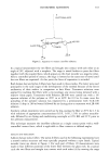
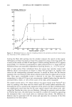
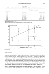
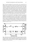
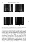
j. Cosmet. Sci., 51, 351-366 (November/December 2000) Measurement of interfiber adhesion Y. K. KAMATH and H.-D. WEIGMANN, TRI/Princeton, P.O. Box 625, Princeton, NJ 08542. Accepted for publication October 25, 2000. Synopsis Two methods of evaluating interfiber adhesive interactions are discussed. A single-point contact method measures the force of separation between two fibers that had been brought into contact with each other at a right angie under a defined contact pressure. After the application of topical treatments, reproducibility of the separation force along the length of the same fiber is poor. This has been attributed to the nonuni- formity of surface deposition and to the uncertainty of the area of contact. The nonuniformity problem disappears when liquids are deposited on the fiber surface. In this case, capillary forces dominate the separation forces, although the uncertainty about the area of real contact between the two fibers still exists. The second method is a multipoint contact method in which the signal is much larger and which represents an average of the adhesive interactions between a single fiber and its neighbors in a hair assembly. The parameter measured is a "withdrawal" or pull-out force from a hair bundle with a defined packing density, which can help in the prediction of compressibility behavior describing hair body. INTRODUCTION Interfiber adhesive forces play an important role in fiber assembly behavior such as bending and compression of fiber bundles. When a fiber assembly is subjected to deformation strains, single fibers slip past one another at their points of contact, thus minimizing the extent of deformation of individual fibers. The efficiency with which this is achieved depends on the forces of interfiber friction that arise as a result of adhesion at the points of contact. The force of adhesion between fibers is difficult to measure and is usually quite inac- curate, mainly because of the uncertainty of the conditions at the points of contact. Kul and Smith (1) have made an attempt to measure adhesive interactions between mohair fibers after treating them with various spinning oils to determine which properties of the oils were important for the properties of the yarn. The principle of their method is shown in Figure 1. Fiber A is mounted on the stationary support D and fiber B is mounted underneath fiber A on a bow. The two fibers are brought into contact with each other at an angle of 90 ø. Fiber B is moved downwards until it separates from fiber A. The force required to bend A (like a cantilever) to the position of detachment of B is measured and treated as the force of adhesion between the two fibers. For fibers coated with spinning oils, the force of adhesion in this arrangement should be equal to the product of contact 351
Purchased for the exclusive use of nofirst nolast (unknown)
From: SCC Media Library & Resource Center (library.scconline.org)