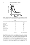
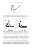
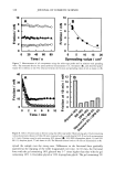
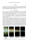
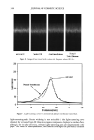
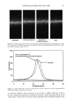
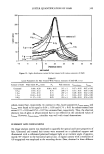
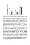
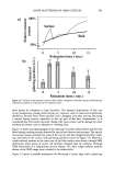
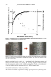
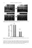
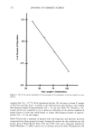
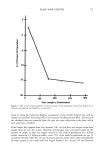
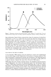
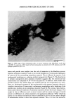
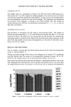
380 JOURNAL OF COSMETIC SCIENCE which in turn, results in improved access to dye molecules, and can be characterized by the diffusion coefficient. Therefore, the diffusion coefficient of a selected dye molecule (under specific dyeing conditions) can be used as a quantitative measure of the damage inflicted upon the fiber. Such methods are used in characterizing changes in the structure of synthetic fibers subjected to different processing methods (1). This study investigates the effects of chemical and photochemical oxidative processes on (a) the microstructure of human hair and (b) the proteins from different histological components of hair. Using microfluorometry, oxidative damage is characterized and quan- tified by studying the changes in diffusion kinetics of the fluorochrome uranine into the hair shaft. The results of the UV radiation-induced photo-oxidative damage to the keratin structure are compared with those obtained in our earlier studies (2) with chemically bleached hair. Increased dye diffusion rates are indicative of changes in fiber morphology. An electrophoretic separation technique was used to show how the main classes of proteins of unaltered hair are modified by cosmetic chemical treatments, light exposure, and com- binations of these processes. The molecular weights of the extractable main classes of proteins of unaltered as well as chemically and photochemically altered hair were es- tablished. Decreases or increases in the amounts of extractable proteins relative to untreated hair suggest which proteins were modified by these chemical/photochemical treatments. Occurrence of new protein bands not observed in untreated hair is indicative of the treatment-induced breakdown of proteins, which were originally not extractable. On the other hand, the absence of protein bands in treated hair, which were observed in untreated hair, suggests further crosslinking of the protein network, which makes them less soluble and therefore less extractable. EXPERIMENTAL HAIR SAMPLES Dark brown hair from DeMeo Brothers, New York, was used. CHEMICAL OXIDATION Bleaching was carried out by two different methods. In one method, hair was bleached for one and four hours with 6% alkaline hydrogen peroxide at room temperature (the pH was adjusted to 10.2 with ammonium hydroxide). The bleaching solution was freshly replaced every 30 minutes. In the other method, hair fibers were treated for 30 minutes with a bleach cream containing hydrogen peroxide and ammonium persulfate (pH 10.2). All samples were thoroughly rinsed, air-dried, and then placed in a desiccator for 24 hours. PHOTOCHEMICAL OXIDATION--EXPOSURE TO SOLAR-SIMULATED UV RADIATION Individual hair fibers were mounted in parallel on templates and exposed for a total of 100,200, 300, 500, and 600 hours to alternating three-hour cycles of UV radiation and humidification as in a QUV accelerated weathering tester. This unit simulates the sunlight in the range of 290-400 nm, with an irradiance maximum at 340 nm. The

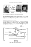
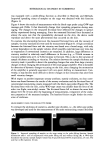
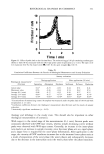
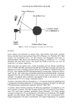
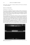
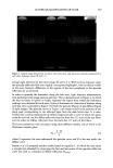
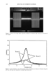
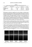
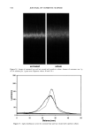
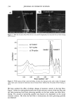
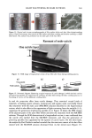
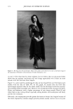
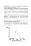
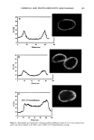
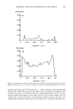
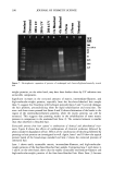
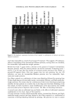
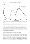
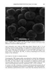
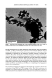
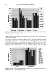
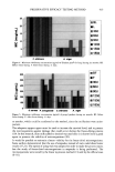
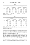
CHEMICAL AND PHOTO-OXIDATIVE HAIR DAMAGE 381 irradiance intensity factor was chosen to be 1.35, compared to 1.0 for regular sunlight. The energy density at the 340-nm wavelength was kept constant at 0.96 W/m 2. The total energy density in the wavelength range of 300-400 nm is 5.06 W/m 2. MICROFLUOROMETRIC STUDY Dyeing with uranine. The photo-oxidized hair fibers as well as the untreated controls were dyed in a 0.1% aqueous uranine solution (pH 7) at 50øC for 5.5 hours. The dyed fibers were rinsed thoroughly for several minutes in warm, distilled water, air-dried, and placed in a desiccator over P205 for 24 hours. Sample preparation for microfluorometry. The dried, dyed fibers were embedded in Spurr's low viscosity resin, cured for 24 hours at 70øC, microtomed at 10-1•m thickness, and viewed in a Leitz MPV 1. ! microspectrophotometer with the Ploem vertical illuminator in the narrow-band blue excitation beam. Spectral scans were obtained on longitudinally viewed fibers, and the wavelength of maximum fluorescence (k,•) was established at 540 nm. Spatial scans were made across the 10-pm-thick fiber cross sections at the established k,, (540 nm). Instrumental settings for spectral and cross-sectional scans ß Blue excitation: 450•490 nm KP: 510 nm LP: 515 nm ß Objective: 25x for spectral scans 40x for cross-sectional scans ß Wavelength, k,,: 540 nm for cross-sectional scans ß Measuring sensor: 20 x 30 pm 2 for spectral scans 3.1 x 25.0 pm 2 for cross-sectional scans ß Accelerating voltage: 1.6 kv ß Scanning speed: 7.2 pm/s Micrographs of the cross-sectional views were made with Kodak slide film at 160 ASA at 60-seconds exposure time, using a 25x objective, 1.6x collar, and a 10x ocular). ELECTROPHORETIC STUDY Hair samples/treatments. Hair samples with treatment sequences were: ß Untreated ß Untreated 100 h/P00 h/300 h UV ß Bleached 1 h/4 h with alkaline 6% H202 ß Bleached 1 h/4 h as above 100 h/300 h UV ß Bleached 1 h/4 h H202/300 h UV/bleached 1 h H202 ß Permed lx/3x ß Permed 1 x/3x as above 100 h/300 h UV ß Bleached/permed ß Bleached/permed/300 h UV Extraction of hair proteins. From each of the above listed categories, 5 to 10 mg of 5-mm-long hair segments were immersed in an extraction buffer containing 0.05 M dithiothreitol (DTT) as reductant, 8 M urea as denaturing agent, and 0.05 M Tris (hydroxymethyl) amino-methane. The ratio of hair to extraction buffer was 1:100. The samples were extracted for 24 hours at ambient temperature, and finally sonificated for 30 minutes. The following reaction with DTT helps to open the keratin matrix to
Purchased for the exclusive use of nofirst nolast (unknown)
From: SCC Media Library & Resource Center (library.scconline.org)