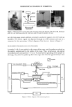
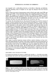
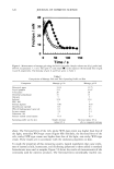
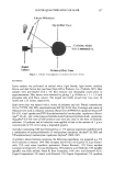
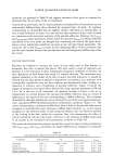
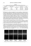
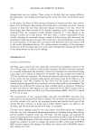
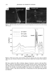
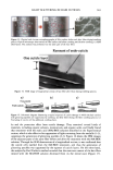
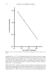
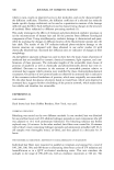
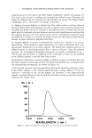
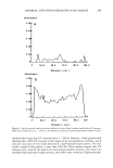
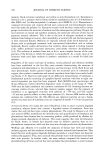
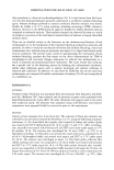
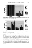
398 JOURNAL OF COSMETIC SCIENCE washing with water:methanol (1:1) and four times with water, the black pellet was again treated with protease and DTT as described in step 3. The final pigment collected by centrifugation was dried over NaOH overnight and equilibrated with saturated aqueous CaC12 for 24 hr. The final yield of melanin was 600 mg from 15 g of Asian black hair (4%). Environmental scanning electron microscopy stz/dies (ESEM). ESEM studies were conducted using the FEI ESEM (model E-3) equipped with a lanthanum hexaboride (LAB6) fila- ment. The microscope was equipped with a Peltier cooling stage (used to maintain the moist environment), which was operated at 2.0ø-3.0øC while the chamber pressure was maintained between 4 and 5 Torr. The sample chamber was pumped down to 5 Torr and then increased to 9 Torr. This was repeated eight times while maintaining a Peltier temperature of 2ø-3øC. Samples were then viewed at 10-24 kV while maintaining a chamber pressure of 4-5 Tort. Occasionally when the liquid precluded the viewing of melanin, the pressure was decreased to remove surface liquid and reveal the melanin. The ESEM was also equipped with the Image Acquisition and Archive System (IAAS), which allowed the results to be recorded and stored digitally as TIF files. Transmission electron microscopy studies (TEM). A Jeol 1200-EX transmission electron mi- croscope equipped with a tungsten filament was used for all imaging studies. The vacuum in the chamber was maintained at 10 3 Pa. Samples were viewed at 100 keV with a low beam current of 63-65 uA. An AMT digital camera system was attached to the port of the TEM. The images were captured, and the signal was sent to a PC for image storage and analysis. The specimen support grids used in the experiments were 200-mesh gold finder grids. Finder grids were used to provide an ease of relocation in interesting areas of the grid. The gold grids had a formvar support film and a carbon coat. The specimens were observed under the following sample preparation: A 5-1nl drop of melanosome suspension was placed on a formvar/carbon-coated gold grid. The sample was allowed to settle for one minute, excess liquid was gently drained off with filter paper, and the sample was allowed to dry. UV-Vis and fluorescence spectroscopic studies. UV-Vis spectral studies were performed using a Shimadzu Multispec-1501 UV-Vis spectrophotometer with diode array detection. Time-dependent spectral measurements were recorded in the scan mode. The fluores- cence studies were performed using a Cary Eclipse fluorescence spectrophotometer. RESULTS UV-VIS ABSORPTION PROPERTIES OF HAIR AND SEPIA MELANIN The absorption spectra of aqueous suspensions (0.1 mg/ml) of hair and Sepia melanin at neutral pH are shown in Figure 1. The absorption of hair melanin exhibits a structureless spectrum with absorptivity decreasing monotonically with decreasing wavelength. Sepia melanin exhibits a higher absorption at longer wavelengths, indicating that the particles are bigger than that of hair melanin. The longer wavelength absorption is attributed to scattering and absorption due to higher-molecular-weight melanin polymers. It is true that the spectral features of melanin change with size and that a sonicated suspension of hair melanin filtered through a micron-size filter displays an absorption spectrum with a decrease in longer wavelength absorptivity (data not shown).

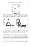
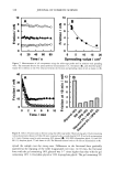
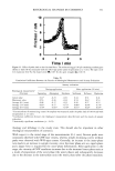
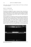
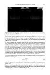
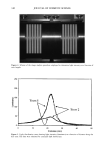
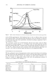
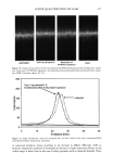
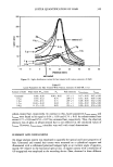
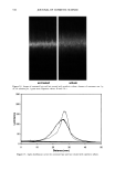
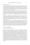
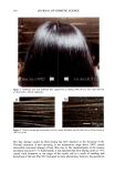
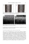
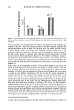
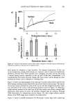
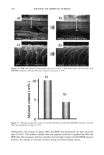
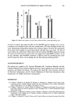
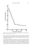
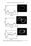
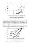
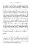
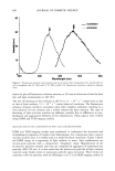
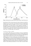
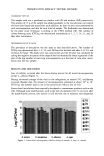
AMMONIA/PEROXIDE BLEACHING OF HAIR 399 D.?• - Figure l. Absorption spectra of aqueous suspensions of hair and Sepia melanin at pH 7.0. Sonicated suspensions of 0.1 mg/ml of melanosomes in water were used for the absorption spectral measurements. FLUORESCENCE PROPERTIES OF HAIR AND SEPIA MELANIN Both isolated hair melanosomes and Sepia melanin at pH 10.0 did not show any fluo- rescence before bleaching. However, after bleaching with NH 3 and H202 at pH 10, both melanins exhibited fluorescence properties. Figure 2 shows the fluorescence emission (}tex = 350 rim) and excitation spectra (}ter n = 450 rim) of hair melanin (0.5 mg/ml) after 45 min of treatment with 2% H202 and 0.75% NH 3 at pH 10.0. The emission maximum is at 468 nm for any excitation wavelength from 300-350 nm. No other emission band is observed for hair melanin. These results agree with the finding by Kayatz et aL (18) that both synthetic melanin and isolated bovine melanosomes fluoresce only after oxidation. However, the fluorescence emission band is reported to be at 548 nm for bovine melanosomes, while hair melanosomes did not show any fluorescence emission in this region. Figure 3 shows the emission and excitation spectra of Sepia melanin under identical conditions of treatment with ammonia and peroxide. The fluorescence emission spec- trum of Sepia melanin showed a maximum at 460 nm (}tex = 350 rim) and an excitation maximum at 349 nm (}tern = 450 rim). The excitation spectra of both Sepia and hair melanin exhibited maxima around the 350 nm region, suggesting that a yellow chro- mophore is responsible for the fluorescence emission at 460 nm. The rates of ammonia/peroxide bleaching of both Sepia and hair melanins were studied by time-dependent fluorescence measurements under identical conditions. Suspensions of 0.2 mg/ml of melanin samples were treated with 1% NH 3 and 2% H202 at pH 10.0 and 25øC. Oxidation of melanins results in fluorescence emission at 450 nm, as shown above. The intensity of fluorescence emission increases with increase in time. Figure 4
Purchased for the exclusive use of nofirst nolast (unknown)
From: SCC Media Library & Resource Center (library.scconline.org)