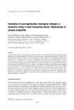
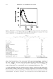
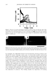
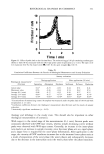
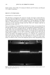
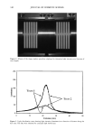
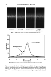
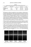
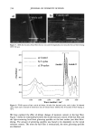
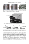
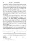
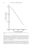
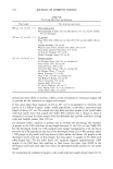
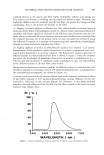
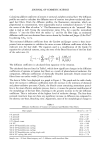
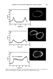
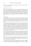
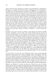
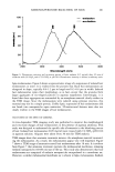
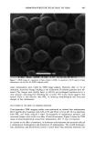
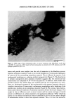
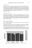
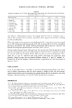
400 JOURNAL OF COSMETIC SCIENCE 3O 20 10 ø / 200 300 excitation ..-' \ ! \ i Wavelenõth {rim} Figure 2. Fluorescence emission and excitation spectra of isolated hair melanosomes (0.5 mg/ml) after 45 min of oxidation with 2% H202 and 0.75% NH3 at pH 10.0. Fluorescence intensity is shown in arbitrary units. shows the plot of fluorescence emission intensity at 450 nm as a function of time for both hair and Sepia melanosomes at pH 10.0. The rate of bleaching of hair melanin at pH 10 is 2.1 x 10 -4 s -1, nearly twice as fast as that of Sepia melanin, 1.2 x 10 -4 s -1, under identical conditions. The fluorescence emission intensity reached a saturation value after complete oxidation, resulting in a clear solution for hair melanin and a yellow solution for Sepia melanin. The rates of bleaching of Sepia and hair melanin are different, possibly due to differences in mor- phological and aggregation behavior of the melanosomes. These aspects were verified using ESEM and TEM imaging studies. ESEM AND TEM STUI)IES: COMPARISON OF SEPIA AND HAIR MELANOSOMES ESEM and TEM imaging studies were performed to understand the structural and morphological properties of isolated hair melanosomes. For comparison, Sepia melanin was also studied, since it is widely used as a model for black eumelanin. Figure 5 shows the ESEM image of a suspension of Sepia melanin in water. Sepia melanosomes are micron-sized particles with a characteristic "doughnut" shape. Magnification of the micron-size particles revealed that they are comprised of aggregates of nanometer-size particles (100-150 nm). It is also noted that the micron-size particles of Sepia melanin could be easily broken down to nanoscopic particles upon sonication. These results are consistent with the previous work by Nofsinger eta/. (11) on SEM imaging studies of

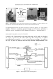
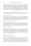
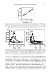
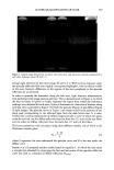
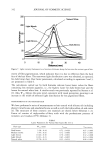
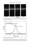
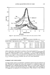
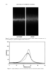
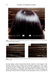
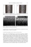
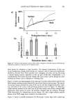
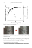
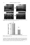
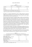
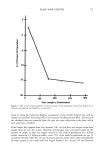
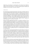
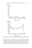
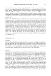
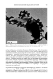
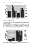
AMMONIA/PEROXIDE BLEACHING OF HAIR 401 1 oo 80 • 60 40 20 -- emission ........ excitation 0 I I I I 200 300 400 500 600 Wavelength (nm) Figure 3. Fluorescence emission and excitation spectra of Sepia melanin (0.5 mg/ml) after 45 min of oxidation with 2% H202 and 0.75% NH 3 at pH 10.0. Fluorescence intensity is shown in arbitrary units. Sepia melanosomes. Figure 6 shows a representative image of a suspension of isolated hair melanosomes in water. It is evident from the pictures that black hair melanosomes are elongated in shape, typically 0.8-1.2 lam in length and 0.3-0.6 lam in width. Isolated hair melanosornes retain their morphology, as in hair, except that the granules form larger aggregates of rice-shaped particles in aqueous suspensions. Interestingly, it is found that these aggregates are surrounded by an amorphous material clearly evident in the TEM image. Since the melanosomes were isolated using protease enzymes, this material may not be a simple protein. Unlike Sepia, suspension of hair melanosomes did not break into nanoparticles upon sonication. Ultrastructural features were also not clearly evident in the TEM images of hair melanosomes. TEM STUDIES ON THE EFFECT OF AMMONIA A time-dependent TEM imaging study was performed to monitor the morphological and structural changes of hair melanosomes in the presence of aqueous ammonia. This study was designed to understand the specific role of ammonia in the bleaching process of hair. Isolated hair melanosomes (0.05 mg/ml) were treated with 1% NH 3 (pH 10.0) in aqueous solution. Aliquots were taken every 30 min for TEM analysis. TEM images show that ammonia treatment removes the amorphous material surround- ing the melanosomes. As a result, the melanosomes appear very well dispersed. Figure 7 shows a TEM image of ammonia-treated hair melanosomes after 30 min. It is evident from Figure 7 that ammonia treatment ruptures the melanosomal membrane, re!eaging melanin nanoparticles (30-60 nm) out of the sac. This study also demonstrates that hair melanosomes are comprised of melanin nanoparticles encapsulated in a membrane sac. However, a similar melanosomal membrane sac is absent in Sepia melanosomes. Sodium
Purchased for the exclusive use of nofirst nolast (unknown)
From: SCC Media Library & Resource Center (library.scconline.org)